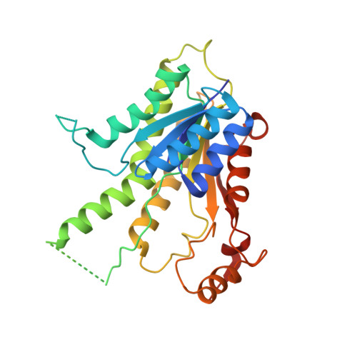
2BF7

Leishmania major pteridine reductase 1 in complex with NADP and biopterin


Experimental Data Snapshot

  • Method: X-RAY DIFFRACTION
  • Resolution: 2.40 Å
  • R-Value Free: 0.242 
  • R-Value Work: 0.219 
  • R-Value Observed: 0.220 

Starting Model: experimental
View more details

wwPDB Validation   3D Report Full Report


Ligand Structure Quality Assessment 


This is version 1.4 of the entry. See complete history


Literature

Structures of Leishmania Major Pteridine Reductase Complexes Reveal the Active Site Features Important for Ligand Binding and to Guide Inhibitor Design

Schuettelkopf, A.W.Hardy, L.W.Beverley, S.M.Hunter, W.N.

(2005) J Mol Biol 352: 105

  • DOI: https://doi.org/10.1016/j.jmb.2005.06.076
  • Primary Citation of Related Structures:  
    2BF7, 2BFA, 2BFM, 2BFO, 2BFP

  • PubMed Abstract: 

    Pteridine reductase (PTR1) is an NADPH-dependent short-chain reductase found in parasitic trypanosomatid protozoans. The enzyme participates in the salvage of pterins and represents a target for the development of improved therapies for infections caused by these parasites. A series of crystallographic analyses of Leishmania major PTR1 are reported. Structures of the enzyme in a binary complex with the cofactor NADPH, and ternary complexes with cofactor and biopterin, 5,6-dihydrobiopterin, and 5,6,7,8-tetrahydrobiopterin reveal that PTR1 does not undergo any major conformational changes to accomplish binding and processing of substrates, and confirm that these molecules bind in a single orientation at the catalytic center suitable for two distinct reductions. Ternary complexes with cofactor and CB3717 and trimethoprim (TOP), potent inhibitors of thymidylate synthase and dihydrofolate reductase, respectively, have been characterized. The structure with CB3717 reveals that the quinazoline moiety binds in similar fashion to the pterin substrates/products and dominates interactions with the enzyme. In the complex with TOP, steric restrictions enforced on the trimethoxyphenyl substituent prevent the 2,4-diaminopyrimidine moiety from adopting the pterin mode of binding observed in dihydrofolate reductase, and explain the inhibition properties of a range of pyrimidine derivates. The molecular detail provided by these complex structures identifies the important interactions necessary to assist the structure-based development of novel enzyme inhibitors of potential therapeutic value.


  • Organizational Affiliation

    Division of Biological Chemistry and Molecular Microbiology, School of Life Sciences, University of Dundee, Dundee DD1 5EH, UK.


Macromolecules
Find similar proteins by:  (by identity cutoff)  |  3D Structure
Entity ID: 1
MoleculeChains Sequence LengthOrganismDetailsImage
PTERIDINE REDUCTASE 1
A, B, C, D
288Leishmania majorMutation(s): 0 
EC: 1.5.1.33
UniProt
Find proteins for Q01782 (Leishmania major)
Explore Q01782 
Go to UniProtKB:  Q01782
Entity Groups  
Sequence Clusters30% Identity50% Identity70% Identity90% Identity95% Identity100% Identity
UniProt GroupQ01782
Sequence Annotations
Expand
  • Reference Sequence
Small Molecules
Ligands 3 Unique
IDChains Name / Formula / InChI Key2D Diagram3D Interactions
NAP
Query on NAP

Download Ideal Coordinates CCD File 
E [auth A],
H [auth B],
K [auth C],
O [auth D]
NADP NICOTINAMIDE-ADENINE-DINUCLEOTIDE PHOSPHATE
C21 H28 N7 O17 P3
XJLXINKUBYWONI-NNYOXOHSSA-N
HBI
Query on HBI

Download Ideal Coordinates CCD File 
F [auth A],
I [auth B],
L [auth C],
P [auth D]
7,8-DIHYDROBIOPTERIN
C9 H13 N5 O3
FEMXZDUTFRTWPE-DZSWIPIPSA-N
EDO
Query on EDO

Download Ideal Coordinates CCD File 
G [auth A],
J [auth B],
M [auth C],
N [auth C],
Q [auth D]
1,2-ETHANEDIOL
C2 H6 O2
LYCAIKOWRPUZTN-UHFFFAOYSA-N
Experimental Data & Validation

Experimental Data

  • Method: X-RAY DIFFRACTION
  • Resolution: 2.40 Å
  • R-Value Free: 0.242 
  • R-Value Work: 0.219 
  • R-Value Observed: 0.220 
  • Space Group: P 21 21 21
Unit Cell:
Length ( Å )Angle ( ˚ )
a = 95.269α = 90
b = 104.554β = 90
c = 138.069γ = 90
Software Package:
Software NamePurpose
REFMACrefinement
DENZOdata reduction
SCALEPACKdata scaling
AMoREphasing

Structure Validation

View Full Validation Report


Ligand Structure Quality Assessment 


Entry History 

Deposition Data

Revision History  (Full details and data files)

  • Version 1.0: 2005-08-31
    Type: Initial release
  • Version 1.1: 2011-05-08
    Changes: Version format compliance
  • Version 1.2: 2011-07-13
    Changes: Version format compliance
  • Version 1.3: 2019-03-06
    Changes: Data collection, Experimental preparation, Other
  • Version 1.4: 2023-12-13
    Changes: Data collection, Database references, Other, Refinement description