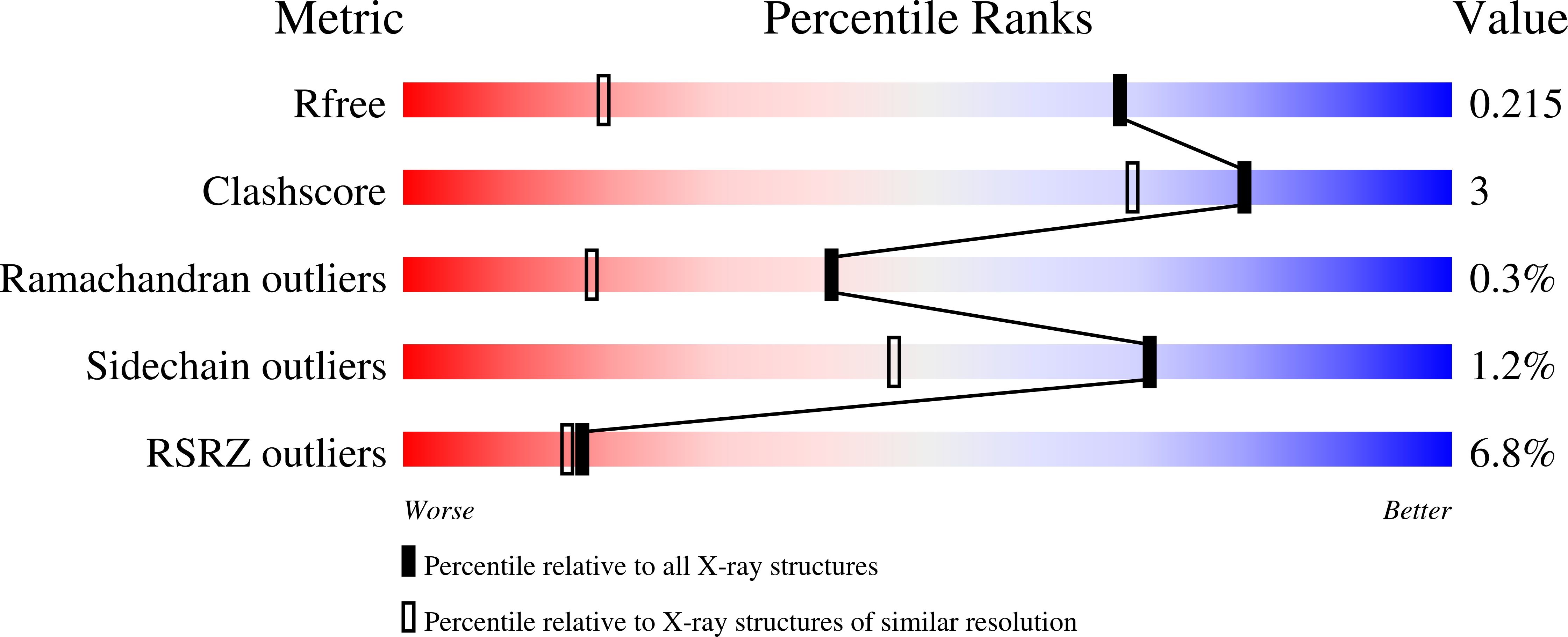
2GCO

Crystal structure of the human RhoC-GppNHp complex


Experimental Data Snapshot

  • Method: X-RAY DIFFRACTION
  • Resolution: 1.40 Å
  • R-Value Free: 0.219 
  • R-Value Work: 0.197 
  • R-Value Observed: 0.198 

Starting Model: experimental
View more details

wwPDB Validation   3D Report Full Report


Ligand Structure Quality Assessment 


This is version 1.3 of the entry. See complete history


Literature

X-ray Crystal Structures Reveal Two Activated States for RhoC.

Dias, S.M.G.Cerione, R.A.

(2007) Biochemistry 46: 6547-6558

  • DOI: https://doi.org/10.1021/bi700035p
  • Primary Citation of Related Structures:  
    2GCN, 2GCO, 2GCP

  • PubMed Abstract: 

    RhoC is a member of the Rho family of Ras-related (small) GTPases and shares significant sequence similarity with the founding member of the family, RhoA. However, despite their similarity, RhoA and RhoC exhibit different binding preferences for effector proteins and give rise to distinct cellular outcomes, with RhoC being directly implicated in the invasiveness of cancer cells and the development of metastasis. While the structural analyses of the signaling-active and -inactive states of RhoA have been performed, thus far, the work on RhoC has been limited to an X-ray structure for its complex with the effector protein, mDia1 (for mammalian Diaphanous 1). Therefore, in order to gain insights into the molecular basis for RhoC activation, as well as clues regarding how it mediates distinct cellular responses relative to those induced by RhoA, we have undertaken a structural comparison of RhoC in its GDP-bound (signaling-inactive) state versus its GTP-bound (signaling-active) state as induced by the nonhydrolyzable GTP analogues, guanosine 5'-(beta,gamma-iminotriphosphate) (GppNHp) and guanosine 5'-(3-O-thiotriphosphate) (GTPgammaS). Interestingly, we find that GppNHp-bound RhoC only shows differences in its switch II domain, relative to GDP-bound RhoC, whereas GTPgammaS-bound RhoC exhibits differences in both its switch I and switch II domains. Given that each of the nonhydrolyzable GTP analogues is able to promote the binding of RhoC to effector proteins, these results suggest that RhoC can undergo at least two conformational transitions during its conversion from a signaling-inactive to a signaling-active state, similar to what has recently been proposed for the H-Ras and M-Ras proteins. In contrast, the available X-ray structures for RhoA suggest that it undergoes only a single conformational transition to a signaling-active state. These and other differences regarding the changes in the switch domains accompanying the activation of RhoA and RhoC provide plausible explanations for the functional specificity exhibited by the two GTPases.


  • Organizational Affiliation

    Department of Molecular Medicine, College of Veterinary Medicine, Cornell University, Ithaca, New York 14853, USA.


Macromolecules
Find similar proteins by:  (by identity cutoff)  |  3D Structure
Entity ID: 1
MoleculeChains Sequence LengthOrganismDetailsImage
Rho-related GTP-binding protein RhoC201Homo sapiensMutation(s): 1 
Gene Names: RHOCARH9ARHC
UniProt & NIH Common Fund Data Resources
Find proteins for P08134 (Homo sapiens)
Explore P08134 
Go to UniProtKB:  P08134
PHAROS:  P08134
GTEx:  ENSG00000155366 
Entity Groups  
Sequence Clusters30% Identity50% Identity70% Identity90% Identity95% Identity100% Identity
UniProt GroupP08134
Sequence Annotations
Expand
  • Reference Sequence
Find similar proteins by:  (by identity cutoff)  |  3D Structure
Entity ID: 2
MoleculeChains Sequence LengthOrganismDetailsImage
Rho-related GTP-binding protein RhoC201Homo sapiensMutation(s): 0 
Gene Names: RHOCARH9ARHC
UniProt & NIH Common Fund Data Resources
Find proteins for P08134 (Homo sapiens)
Explore P08134 
Go to UniProtKB:  P08134
PHAROS:  P08134
GTEx:  ENSG00000155366 
Entity Groups  
Sequence Clusters30% Identity50% Identity70% Identity90% Identity95% Identity100% Identity
UniProt GroupP08134
Sequence Annotations
Expand
  • Reference Sequence
Small Molecules
Modified Residues  1 Unique
IDChains TypeFormula2D DiagramParent
CSX
Query on CSX
A
L-PEPTIDE LINKINGC3 H7 N O3 SCYS
Experimental Data & Validation

Experimental Data

  • Method: X-RAY DIFFRACTION
  • Resolution: 1.40 Å
  • R-Value Free: 0.219 
  • R-Value Work: 0.197 
  • R-Value Observed: 0.198 
  • Space Group: P 21 21 2
Unit Cell:
Length ( Å )Angle ( ˚ )
a = 64.092α = 90
b = 75.645β = 90
c = 82.903γ = 90
Software Package:
Software NamePurpose
SCALAdata scaling
MOLREPphasing
REFMACrefinement
PDB_EXTRACTdata extraction
CrystalCleardata reduction
CCP4data scaling

Structure Validation

View Full Validation Report


Ligand Structure Quality Assessment 


Entry History 

Deposition Data

Revision History  (Full details and data files)

  • Version 1.0: 2007-03-27
    Type: Initial release
  • Version 1.1: 2008-05-01
    Changes: Version format compliance
  • Version 1.2: 2011-07-13
    Changes: Advisory, Version format compliance
  • Version 1.3: 2023-08-30
    Changes: Data collection, Database references, Derived calculations, Refinement description