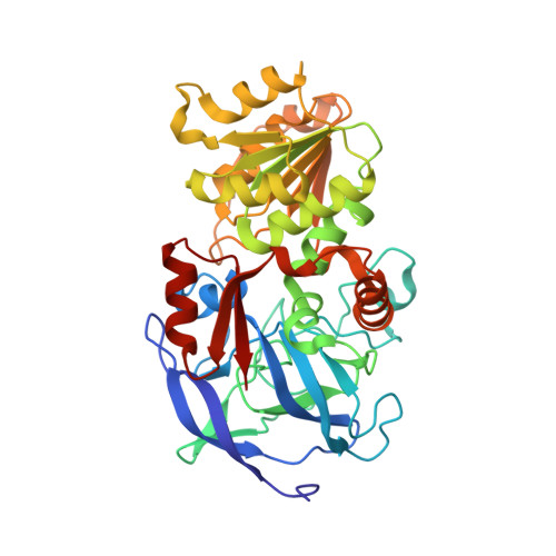
2JHG

Structural evidence for a ligand coordination switch in liver alcohol dehydrogenase


Experimental Data Snapshot

  • Method: X-RAY DIFFRACTION
  • Resolution: 1.20 Å
  • R-Value Free: 0.142 
  • R-Value Work: 0.114 
  • R-Value Observed: 0.115 

Starting Model: experimental
View more details

wwPDB Validation   3D Report Full Report


Ligand Structure Quality Assessment 


This is version 1.4 of the entry. See complete history


Literature

Structural Evidence for a Ligand Coordination Switch in Liver Alcohol Dehydrogenase

Meijers, R.Adolph, H.W.Dauter, Z.Wilson, K.S.Lamzin, V.S.Cedergren-Zeppezauer, E.S.

(2007) Biochemistry 46: 5446

  • DOI: https://doi.org/10.1021/bi6023594
  • Primary Citation of Related Structures:  
    2JHF, 2JHG

  • PubMed Abstract: 

    The use of substrate analogues as inhibitors provides a way to understand and manipulate enzyme function. Here we report two 1 A resolution crystal structures of liver alcohol dehydrogenase in complex with NADH and two inhibitors: dimethyl sulfoxide and isobutyramide. Both structures present a dynamic state of inhibition. In the dimethyl sulfoxide complex structure, the inhibitor is caught in transition on its way to the active site using a flash-freezing protocol and a cadmium-substituted enzyme. One inhibitor molecule is partly located in the first and partly in the second coordination sphere of the active site metal. A hydroxide ion bound to the active site metal lies close to the pyridine ring of NADH, which is puckered in a twisted boat conformation. The cadmium ion is coordinated by both the hydroxide ion and the inhibitor molecule, providing structural evidence of a coordination switch at the active site metal ion. The structure of the isobutyramide complex reveals the partial formation of an adduct between the isobutyramide inhibitor and NADH. It provides evidence of the contribution of a shift from the keto to the enol tautomer during aldehyde reduction. The different positions of the inhibitors further refine the knowledge of the dynamics of the enzyme mechanism and explain how the crowded active site can facilitate the presence of a substrate and a metal-bound hydroxide ion.


  • Organizational Affiliation

    European Molecular Biology Laboratory, Hamburg Outstation, c/o DESY, Notkestrasse 85, D-22603 Hamburg, Federal Republic of Germany. rob.meijers@synchrotron-soleil.fr


Macromolecules
Find similar proteins by:  (by identity cutoff)  |  3D Structure
Entity ID: 1
MoleculeChains Sequence LengthOrganismDetailsImage
ALCOHOL DEHYDROGENASE E CHAIN
A, B
374Equus caballusMutation(s): 0 
EC: 1.1.1.1
UniProt
Find proteins for P00327 (Equus caballus)
Explore P00327 
Go to UniProtKB:  P00327
Entity Groups  
Sequence Clusters30% Identity50% Identity70% Identity90% Identity95% Identity100% Identity
UniProt GroupP00327
Sequence Annotations
Expand
  • Reference Sequence
Small Molecules
Ligands 3 Unique
IDChains Name / Formula / InChI Key2D Diagram3D Interactions
NAD
Query on NAD

Download Ideal Coordinates CCD File 
E [auth A],
I [auth B]
NICOTINAMIDE-ADENINE-DINUCLEOTIDE
C21 H27 N7 O14 P2
BAWFJGJZGIEFAR-NNYOXOHSSA-N
IBO
Query on IBO

Download Ideal Coordinates CCD File 
F [auth A],
J [auth B]
2-METHYLPROPANAMIDE
C4 H9 N O
WFKAJVHLWXSISD-UHFFFAOYSA-N
ZN
Query on ZN

Download Ideal Coordinates CCD File 
C [auth A],
D [auth A],
G [auth B],
H [auth B]
ZINC ION
Zn
PTFCDOFLOPIGGS-UHFFFAOYSA-N
Binding Affinity Annotations 
IDSourceBinding Affinity
IBO BindingDB:  2JHG Ki: min: 1.70e+5, max: 2.20e+5 (nM) from 2 assay(s)
Experimental Data & Validation

Experimental Data

  • Method: X-RAY DIFFRACTION
  • Resolution: 1.20 Å
  • R-Value Free: 0.142 
  • R-Value Work: 0.114 
  • R-Value Observed: 0.115 
  • Space Group: P 1
Unit Cell:
Length ( Å )Angle ( ˚ )
a = 50.186α = 77.12
b = 43.798β = 87.44
c = 92.516γ = 108.89
Software Package:
Software NamePurpose
REFMACrefinement
DENZOdata reduction
SCALEPACKdata scaling

Structure Validation

View Full Validation Report


Ligand Structure Quality Assessment 


Entry History 

Deposition Data

Revision History  (Full details and data files)

  • Version 1.0: 2007-04-24
    Type: Initial release
  • Version 1.1: 2011-05-08
    Changes: Version format compliance
  • Version 1.2: 2011-07-13
    Changes: Version format compliance
  • Version 1.3: 2019-07-24
    Changes: Data collection, Derived calculations
  • Version 1.4: 2023-12-13
    Changes: Data collection, Database references, Derived calculations, Refinement description