2OFP

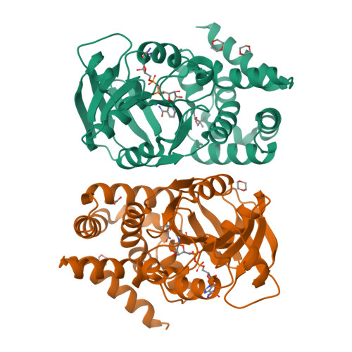
Crystal structure of Escherichia coli ketopantoate reductase in a ternary complex with NADP+ and pantoate


Experimental Data Snapshot

  • Method: X-RAY DIFFRACTION
  • Resolution: 2.30 Å
  • R-Value Free: 0.215 
  • R-Value Work: 0.156 
  • R-Value Observed: 0.159 

Starting Models: experimental
View more details

wwPDB Validation   3D Report Full Report


Ligand Structure Quality Assessment 

Created with Raphaël 2.3.0Worse 01 BetterLigand structure goodness of fit to experimental dataBest fitted NAPClick on this verticalbar to view details

This is version 1.3 of the entry. See complete history


Literature

Crystal Structure of Escherichia coli Ketopantoate Reductase in a Ternary Complex with NADP+ and Pantoate Bound: SUBSTRATE RECOGNITION, CONFORMATIONAL CHANGE, AND COOPERATIVITY.

Ciulli, A.Chirgadze, D.Y.Smith, A.G.Blundell, T.L.Abell, C.

(2007) J Biol Chem 282: 8487-8497

  • DOI: https://doi.org/10.1074/jbc.M611171200
  • Primary Citation of Related Structures:  
    2OFP

  • PubMed Abstract: 

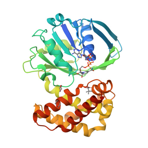
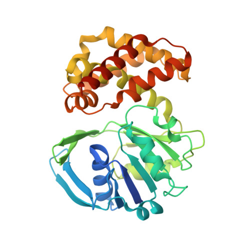
    Ketopantoate reductase (KPR, EC 1.1.1.169) catalyzes the NADPH-dependent reduction of ketopantoate to pantoate, an essential step for the biosynthesis of pantothenate (vitamin B5). Inhibitors of the enzymes of this pathway have been proposed as potential antibiotics or herbicides. Here we present the crystal structure of Escherichia coli KPR in a precatalytic ternary complex with NADP+ and pantoate bound, solved to 2.3 A of resolution. The asymmetric unit contains two protein molecules, each in a ternary complex; however, one is in a more closed conformation than the other. A hinge bending between the N- and C-terminal domains is observed, which triggers the switch of the essential Lys176 to form a key hydrogen bond with the C2 hydroxyl of pantoate. Pantoate forms additional interactions with conserved residues Ser244, Asn98, and Asn180 and with two conservatively varied residues, Asn194 and Asn241. The steady-state kinetics of active site mutants R31A, K72A, N98A, K176A, S244A, and E256A implicate Asn98 as well as Lys176 and Glu256 in the catalytic mechanism. Isothermal titration calorimetry studies with these mutants further demonstrate the importance of Ser244 for substrate binding and of Arg31 and Lys72 for cofactor binding. Further calorimetric studies show that KPR discriminates binding of ketopantoate against pantoate only with NADPH bound. This work provides insights into the roles of active site residues and conformational changes in substrate recognition and catalysis, leading to the proposal of a detailed molecular mechanism for KPR activity.


  • Organizational Affiliation

    University Chemical Laboratory, Department of Biochemistry, University of Cambridge, 80 Tennis Court Road, Cambridge CB2 1GA, United Kingdom.


Macromolecules
Find similar proteins by:  (by identity cutoff)  |  3D Structure
Entity ID: 1
MoleculeChains Sequence LengthOrganismDetailsImage
Ketopantoate reductase
A, B
320Escherichia coli K-12Mutation(s): 0 
Gene Names: panEapbA
EC: 1.1.1.169
UniProt
Find proteins for P0A9J4 (Escherichia coli (strain K12))
Explore P0A9J4 
Go to UniProtKB:  P0A9J4
Entity Groups  
Sequence Clusters30% Identity50% Identity70% Identity90% Identity95% Identity100% Identity
UniProt GroupP0A9J4
Sequence Annotations
Expand
  • Reference Sequence
Small Molecules
Ligands 4 Unique
IDChains Name / Formula / InChI Key2D Diagram3D Interactions
NAP
Query on NAP

Download Ideal Coordinates CCD File 
C [auth A],
H [auth B]
NADP NICOTINAMIDE-ADENINE-DINUCLEOTIDE PHOSPHATE
C21 H28 N7 O17 P3
XJLXINKUBYWONI-NNYOXOHSSA-N
PAF
Query on PAF

Download Ideal Coordinates CCD File 
D [auth A],
I [auth B]
PANTOATE
C6 H11 O4
OTOIIPJYVQJATP-BYPYZUCNSA-M
DIO
Query on DIO

Download Ideal Coordinates CCD File 
E [auth A],
F [auth A],
J [auth B],
K [auth B]
1,4-DIETHYLENE DIOXIDE
C4 H8 O2
RYHBNJHYFVUHQT-UHFFFAOYSA-N
ACT
Query on ACT

Download Ideal Coordinates CCD File 
G [auth B]ACETATE ION
C2 H3 O2
QTBSBXVTEAMEQO-UHFFFAOYSA-M
Experimental Data & Validation

Experimental Data

  • Method: X-RAY DIFFRACTION
  • Resolution: 2.30 Å
  • R-Value Free: 0.215 
  • R-Value Work: 0.156 
  • R-Value Observed: 0.159 
  • Space Group: P 41 21 2
Unit Cell:
Length ( Å )Angle ( ˚ )
a = 101.692α = 90
b = 101.692β = 90
c = 171.168γ = 90
Software Package:
Software NamePurpose
REFMACrefinement
ADSCdata collection
DENZOdata reduction
SCALEPACKdata scaling
AMoREphasing

Structure Validation

View Full Validation Report


Ligand Structure Quality Assessment 

Created with Raphaël 2.3.0Worse 01 BetterLigand structure goodness of fit to experimental dataBest fitted NAPClick on this verticalbar to view details

Entry History 

Deposition Data

Revision History  (Full details and data files)

  • Version 1.0: 2007-01-16
    Type: Initial release
  • Version 1.1: 2007-10-16
    Changes: Version format compliance
  • Version 1.2: 2011-07-13
    Changes: Version format compliance
  • Version 1.3: 2023-08-30
    Changes: Data collection, Database references, Derived calculations, Refinement description