2PDQ

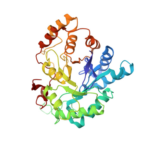
Human aldose reductase mutant C303D complexed with uracil-type inhibitor.


Experimental Data Snapshot

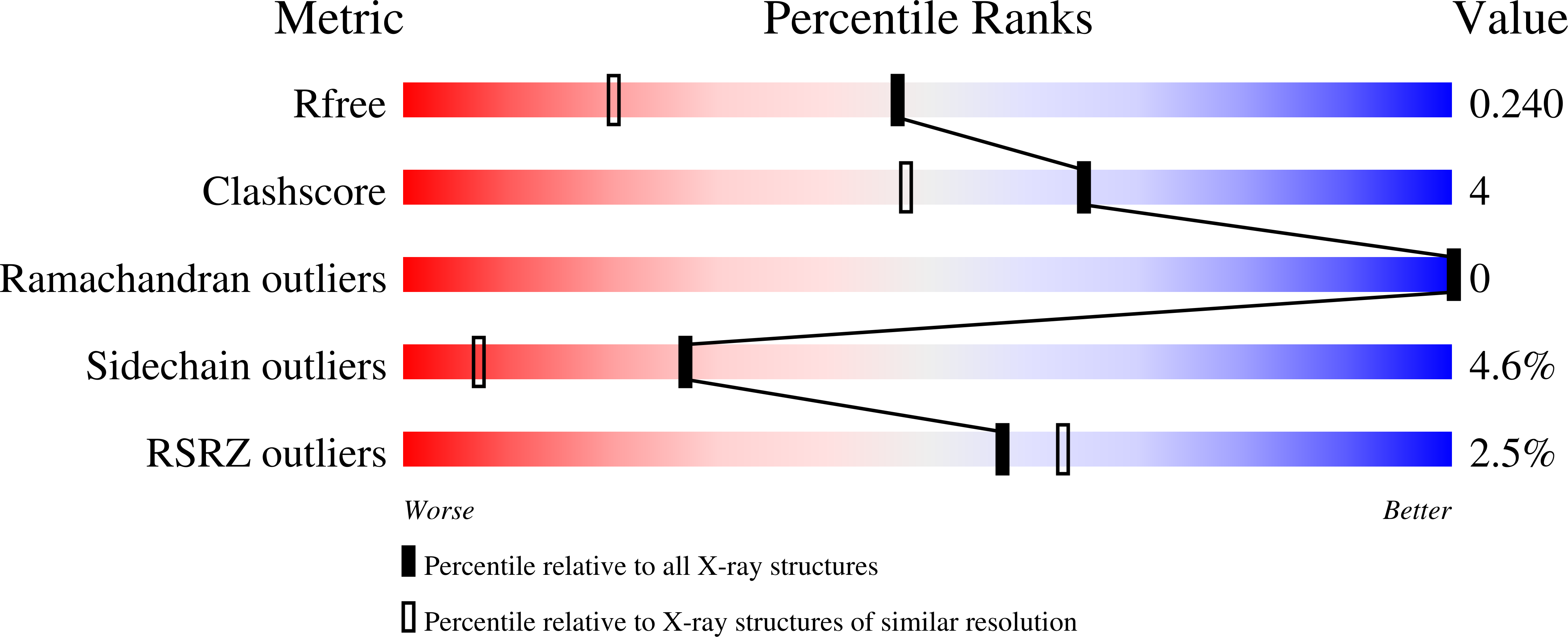
  • Method: X-RAY DIFFRACTION
  • Resolution: 1.73 Å
  • R-Value Free: 0.248 
  • R-Value Work: 0.182 
  • R-Value Observed: 0.184 

Starting Model: experimental
View more details

wwPDB Validation   3D Report Full Report


Ligand Structure Quality Assessment 


This is version 1.3 of the entry. See complete history


Literature

Merging the binding sites of aldose and aldehyde reductase for detection of inhibitor selectivity-determining features.

Steuber, H.Heine, A.Podjarny, A.Klebe, G.

(2008) J Mol Biol 379: 991-1016

  • DOI: https://doi.org/10.1016/j.jmb.2008.03.063
  • Primary Citation of Related Structures:  
    2PD5, 2PD9, 2PDB, 2PDC, 2PDF, 2PDG, 2PDH, 2PDI, 2PDJ, 2PDK, 2PDL, 2PDM, 2PDN, 2PDP, 2PDQ, 2PDU, 2PDW, 2PDX, 2PDY

  • PubMed Abstract: 

    Inhibition of human aldose reductase (ALR2) evolved as a promising therapeutic concept to prevent late complications of diabetes. As well as appropriate affinity and bioavailability, putative inhibitors should possess a high level of selectivity for ALR2 over the related aldehyde reductase (ALR1). We investigated the selectivity-determining features by gradually mapping the residues deviating between the binding pockets of ALR1 and ALR2 into the ALR2 binding pocket. The resulting mutational constructs of ALR2 (eight point mutations and one double mutant) were probed for their influence towards ligand selectivity by X-ray structure analysis of the corresponding complexes and isothermal titration calorimetry (ITC). The binding properties of these mutants were evaluated using a ligand set of zopolrestat, a related uracil derivative, IDD388, IDD393, sorbinil, fidarestat and tolrestat. Our study revealed induced-fit adaptations within the mutated binding site as an essential prerequisite for ligand accommodation related to the selectivity discrimination of the ligands. However, our study also highlights the limits of the present understanding of protein-ligand interactions. Interestingly, binding site mutations not involved in any direct interaction to the ligands in various cases show significant effects towards their binding thermodynamics. Furthermore, our results suggest the binding site residues deviating between ALR1 and ALR2 influence ligand affinity in a complex interplay, presumably involving changes of dynamic properties and differences of the solvation/desolvation balance upon ligand binding.


  • Organizational Affiliation

    Department of Pharmaceutical Chemistry, Philipps-University Marburg, Marbacher Weg 6, 35032 Marburg, Germany.


Macromolecules
Find similar proteins by:  (by identity cutoff)  |  3D Structure
Entity ID: 1
MoleculeChains Sequence LengthOrganismDetailsImage
Aldose reductase316Homo sapiensMutation(s): 1 
Gene Names: ALR2
EC: 1.1.1.21 (PDB Primary Data), 1.1.1.372 (UniProt), 1.1.1.300 (UniProt), 1.1.1.54 (UniProt)
UniProt & NIH Common Fund Data Resources
Find proteins for P15121 (Homo sapiens)
Explore P15121 
Go to UniProtKB:  P15121
PHAROS:  P15121
GTEx:  ENSG00000085662 
Entity Groups  
Sequence Clusters30% Identity50% Identity70% Identity90% Identity95% Identity100% Identity
UniProt GroupP15121
Sequence Annotations
Expand
  • Reference Sequence
Small Molecules
Ligands 2 Unique
IDChains Name / Formula / InChI Key2D Diagram3D Interactions
NAP
Query on NAP

Download Ideal Coordinates CCD File 
B [auth A]NADP NICOTINAMIDE-ADENINE-DINUCLEOTIDE PHOSPHATE
C21 H28 N7 O17 P3
XJLXINKUBYWONI-NNYOXOHSSA-N
47D
Query on 47D

Download Ideal Coordinates CCD File 
C [auth A]{3-[(5-CHLORO-1,3-BENZOTHIAZOL-2-YL)METHYL]-2,4-DIOXO-3,4-DIHYDROPYRIMIDIN-1(2H)-YL}ACETIC ACID
C14 H10 Cl N3 O4 S
RQWICELTTDJODO-UHFFFAOYSA-N
Binding Affinity Annotations 
IDSourceBinding Affinity
47D BindingDB:  2PDQ IC50: 5.7 (nM) from 1 assay(s)
Experimental Data & Validation

Experimental Data

  • Method: X-RAY DIFFRACTION
  • Resolution: 1.73 Å
  • R-Value Free: 0.248 
  • R-Value Work: 0.182 
  • R-Value Observed: 0.184 
  • Space Group: P 1
Unit Cell:
Length ( Å )Angle ( ˚ )
a = 40.32α = 76.07
b = 47.13β = 67.22
c = 47.404γ = 76.89
Software Package:
Software NamePurpose
SHELXmodel building
SHELXL-97refinement
CrystalCleardata collection
HKL-2000data reduction
HKL-2000data scaling
CNSphasing

Structure Validation

View Full Validation Report


Ligand Structure Quality Assessment 


Entry History 

Deposition Data

Revision History  (Full details and data files)

  • Version 1.0: 2008-04-01
    Type: Initial release
  • Version 1.1: 2011-07-13
    Changes: Version format compliance
  • Version 1.2: 2021-10-20
    Changes: Database references, Derived calculations
  • Version 1.3: 2023-08-30
    Changes: Data collection, Refinement description