2QTS | pdb_00002qts

Structure of an acid-sensing ion channel 1 at 1.9 A resolution and low pH


Experimental Data Snapshot

  • Method: X-RAY DIFFRACTION
  • Resolution: 1.90 Å
  • R-Value Free: 
    0.233 (Depositor), 0.210 (DCC) 
  • R-Value Work: 
    0.208 (Depositor) 
  • R-Value Observed: 
    0.208 (Depositor) 

wwPDB Validation   3D Report Full Report


Ligand Structure Quality Assessment 

Created with Raphaël 2.3.0Worse 01 BetterLigand structure goodness of fit to experimental dataBest fitted NAGClick on this verticalbar to view details

This is version 2.1 of the entry. See complete history


Literature

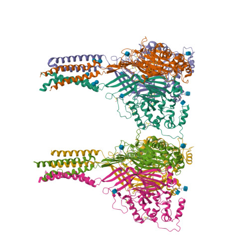
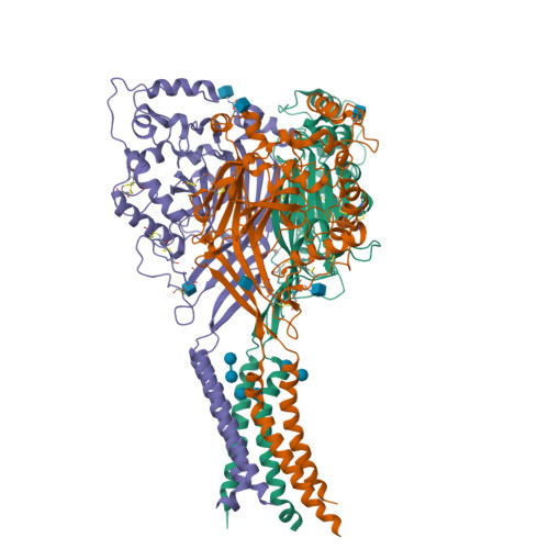
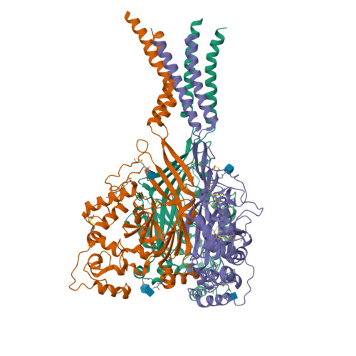
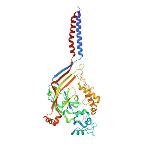
Structure of acid-sensing ion channel 1 at 1.9A resolution and low pH

Jasti, J.Furukawa, H.Gonzales, E.B.Gouaux, E.

(2007) Nature 449: 316-323

  • DOI: https://doi.org/10.1038/nature06163
  • Primary Citation of Related Structures:  
    2QTS

  • PubMed Abstract: 

    Acid-sensing ion channels (ASICs) are voltage-independent, proton-activated receptors that belong to the epithelial sodium channel/degenerin family of ion channels and are implicated in perception of pain, ischaemic stroke, mechanosensation, learning and memory. Here we report the low-pH crystal structure of a chicken ASIC1 deletion mutant at 1.9 A resolution. Each subunit of the chalice-shaped homotrimer is composed of short amino and carboxy termini, two transmembrane helices, a bound chloride ion and a disulphide-rich, multidomain extracellular region enriched in acidic residues and carboxyl-carboxylate pairs within 3 A, suggesting that at least one carboxyl group bears a proton. Electrophysiological studies on aspartate-to-asparagine mutants confirm that these carboxyl-carboxylate pairs participate in proton sensing. Between the acidic residues and the transmembrane pore lies a disulphide-rich 'thumb' domain poised to couple the binding of protons to the opening of the ion channel, thus demonstrating that proton activation involves long-range conformational changes.


  • Organizational Affiliation

    Vollum Institute, Oregon Health and Science University, 3181 SW Sam Jackson Park Road, Portland, Oregon 97239, USA.


Macromolecules
Find similar proteins by:  (by identity cutoff)  |  3D Structure
Entity ID: 1
MoleculeChains Sequence LengthOrganismDetailsImage
Acid-sensing ion channel
A, B, C, D, E
438Gallus gallusMutation(s): 0 
Gene Names: ACCN2
Membrane Entity: Yes 
UniProt
Find proteins for Q1XA76 (Gallus gallus)
Explore Q1XA76 
Go to UniProtKB:  Q1XA76
Entity Groups  
Sequence Clusters30% Identity50% Identity70% Identity90% Identity95% Identity100% Identity
UniProt GroupQ1XA76
Glycosylation
Glycosylation Sites: 2
Sequence Annotations
Expand
  • Reference Sequence
Oligosaccharides

Help

Entity ID: 2
MoleculeChains Length2D Diagram Glycosylation3D Interactions
alpha-D-glucopyranose-(1-4)-alpha-D-glucopyranose
G, H, I
2N/A
Glycosylation Resources
GlyTouCan:  G07411ON
GlyCosmos:  G07411ON
Small Molecules
Ligands 2 Unique
IDChains Name / Formula / InChI Key2D Diagram3D Interactions
NAG
Query on NAG

Download Ideal Coordinates CCD File 
AA [auth F]
J [auth A]
K [auth A]
M [auth B]
N [auth B]
2-acetamido-2-deoxy-beta-D-glucopyranose
C8 H15 N O6
OVRNDRQMDRJTHS-FMDGEEDCSA-N
CL
Query on CL

Download Ideal Coordinates CCD File 
L [auth A]
O [auth B]
R [auth C]
S [auth D]
V [auth E]
CHLORIDE ION
Cl
VEXZGXHMUGYJMC-UHFFFAOYSA-M
Biologically Interesting Molecules (External Reference) 1 Unique
Experimental Data & Validation

Experimental Data

  • Method: X-RAY DIFFRACTION
  • Resolution: 1.90 Å
  • R-Value Free:  0.233 (Depositor), 0.210 (DCC) 
  • R-Value Work:  0.208 (Depositor) 
  • R-Value Observed: 0.208 (Depositor) 
Space Group: P 1 21 1
Unit Cell:
Length ( Å )Angle ( ˚ )
a = 124.252α = 90
b = 110.781β = 97.54
c = 149.891γ = 90
Software Package:
Software NamePurpose
CNSrefinement
PDB_EXTRACTdata extraction
BOSdata collection
HKL-2000data reduction
HKL-2000data scaling
SOLVEphasing

Structure Validation

View Full Validation Report


Ligand Structure Quality Assessment 

Created with Raphaël 2.3.0Worse 01 BetterLigand structure goodness of fit to experimental dataBest fitted NAGClick on this verticalbar to view details

Entry History 

Deposition Data

Revision History  (Full details and data files)

  • Version 1.0: 2007-09-25
    Type: Initial release
  • Version 1.1: 2011-07-13
    Changes: Derived calculations, Version format compliance
  • Version 2.0: 2020-07-29
    Type: Remediation
    Reason: Carbohydrate remediation
    Changes: Atomic model, Data collection, Derived calculations, Non-polymer description, Structure summary
  • Version 2.1: 2024-10-30
    Changes: Data collection, Database references, Structure summary