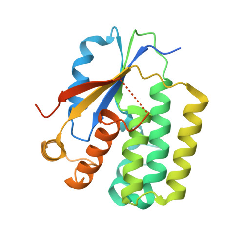
2VPP

Drosophila melanogaster deoxyribonucleoside kinase successfully activates gemcitabine in transduced cancer cell lines


Experimental Data Snapshot

  • Method: X-RAY DIFFRACTION
  • Resolution: 2.20 Å
  • R-Value Free: 0.253 
  • R-Value Work: 0.207 
  • R-Value Observed: 0.209 

Starting Model: experimental
View more details

wwPDB Validation   3D Report Full Report


Ligand Structure Quality Assessment 


This is version 1.3 of the entry. See complete history


Literature

Drosophila Melanogaster Deoxyribonucleoside Kinase Activates Gemcitabine.

Knecht, W.Mikkelsen, N.E.Clausen, A.Willer, M.Eklund, H.Gojkovic, Z.Piskur, J.

(2009) Biochem Biophys Res Commun 382: 430

  • DOI: https://doi.org/10.1016/j.bbrc.2009.03.041
  • Primary Citation of Related Structures:  
    2VPP

  • PubMed Abstract: 

    Drosophila melanogaster multisubstrate deoxyribonucleoside kinase (Dm-dNK) can additionally sensitize human cancer cell lines towards the anti-cancer drug gemcitabine. We show that this property is based on the Dm-dNK ability to efficiently phosphorylate gemcitabine. The 2.2A resolution structure of Dm-dNK in complex with gemcitabine shows that the residues Tyr70 and Arg105 play a crucial role in the firm positioning of gemcitabine by extra interactions made by the fluoride atoms. This explains why gemcitabine is a good substrate for Dm-dNK.


  • Organizational Affiliation

    BioCentrum-DTU, Technical University of Denmark, Lyngby, Denmark.


Macromolecules
Find similar proteins by:  (by identity cutoff)  |  3D Structure
Entity ID: 1
MoleculeChains Sequence LengthOrganismDetailsImage
DEOXYNUCLEOSIDE KINASE
A, B
230Drosophila melanogasterMutation(s): 0 
EC: 2.7.1.145
UniProt
Find proteins for Q9XZT6 (Drosophila melanogaster)
Explore Q9XZT6 
Go to UniProtKB:  Q9XZT6
Entity Groups  
Sequence Clusters30% Identity50% Identity70% Identity90% Identity95% Identity100% Identity
UniProt GroupQ9XZT6
Sequence Annotations
Expand
  • Reference Sequence
Small Molecules
Experimental Data & Validation

Experimental Data

  • Method: X-RAY DIFFRACTION
  • Resolution: 2.20 Å
  • R-Value Free: 0.253 
  • R-Value Work: 0.207 
  • R-Value Observed: 0.209 
  • Space Group: P 21 21 2
Unit Cell:
Length ( Å )Angle ( ˚ )
a = 120.106α = 90
b = 62.298β = 90
c = 68.078γ = 90
Software Package:
Software NamePurpose
REFMACrefinement
MOSFLMdata reduction
SCALAdata scaling
PHASERphasing

Structure Validation

View Full Validation Report


Ligand Structure Quality Assessment 


Entry History 

Deposition Data

Revision History  (Full details and data files)

  • Version 1.0: 2009-03-24
    Type: Initial release
  • Version 1.1: 2011-05-08
    Changes: Version format compliance
  • Version 1.2: 2011-07-13
    Changes: Version format compliance
  • Version 1.3: 2023-12-13
    Changes: Data collection, Database references, Derived calculations, Other, Refinement description