2WZE

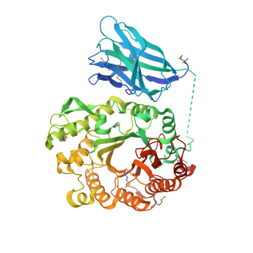
High resolution crystallographic structure of the Clostridium thermocellum N-terminal endo-1,4-beta-D-xylanase 10B (Xyn10B) CBM22-1- GH10 modules complexed with xylohexaose


Experimental Data Snapshot

  • Method: X-RAY DIFFRACTION
  • Resolution: 2.50 Å
  • R-Value Free: 0.189 
  • R-Value Work: 0.147 
  • R-Value Observed: 0.149 

wwPDB Validation   3D Report Full Report


This is version 2.1 of the entry. See complete history


Literature

Putting an N-Terminal End to the Clostridium Thermocellum Xylanase Xyn10B Story: Crystal Structure of the Cbm22-1-Gh10 Modules Complexed with Xylohexaose.

Najmudin, S.Pinheiro, B.A.Prates, J.A.M.Gilbert, H.J.Romao, M.J.Fontes, C.M.G.A.

(2010) J Struct Biol 172: 353

  • DOI: https://doi.org/10.1016/j.jsb.2010.07.009
  • Primary Citation of Related Structures:  
    2W5F, 2WYS, 2WZE

  • PubMed Abstract: 

    In general, plant cell wall degrading enzymes are modular proteins containing catalytic domains linked to one or more non-catalytic carbohydrate-binding modules (CBMs). Xyn10B from Clostridium thermocellum is a typical modular enzyme containing an N-terminal family 22 CBM (CBM22-1), a family 10 glycoside hydrolase catalytic domain (GH10), a second CBM22 (CBM22-2), a dockerin sequence and a C-terminal family 1 carbohydrate esterase (CE1) catalytic domain. The structure of the N-terminal bi-modular CBM22-1-GH10 component of Xyn10B has been determined using a SeMet derivative by SAD to 2.5Å. The data was extended to 2.0Å for the non-SeMet mutant complexed with xylohexaose. CBM22-1-GH10 is a 60kDa protein with an E337A mutation to render the GH10 subunit inactive. Three of the six xylose residues of xylohexaose are shown to be bound in the inactivated GH10 substrate binding cleft, with the other three sugars presumably disordered in the solvent channel. The protein is a dimer in the asymmetric unit with extensive surface contacts between the two GH10 modules and between the CBM22-1 and GH10 modules. Residues from helix H4 of the GH10 module provide the major contacts by fitting into the minor groove of the CBM22-1 module. The orientation of CBM22-1 is such that it would allow the substrate to be loosely bound and subsequently delivered to the active site in a processive manner.


  • Organizational Affiliation

    CIISA - Faculdade de Medicina Veterinária, Universidade Técnica de Lisboa, Avenida da Universidade Técnica, 1300-477 Lisboa, Portugal. shabir@fmv.utl.pt


Macromolecules
Find similar proteins by:  (by identity cutoff)  |  3D Structure
Entity ID: 1
MoleculeChains Sequence LengthOrganismDetailsImage
ENDO-1,4-BETA-XYLANASE Y
A, B
540Acetivibrio thermocellusMutation(s): 1 
EC: 3.2.1.8
UniProt
Find proteins for P51584 (Acetivibrio thermocellus)
Explore P51584 
Go to UniProtKB:  P51584
Entity Groups  
Sequence Clusters30% Identity50% Identity70% Identity90% Identity95% Identity100% Identity
UniProt GroupP51584
Sequence Annotations
Expand
  • Reference Sequence
Oligosaccharides

Help

Entity ID: 2
MoleculeChains Length2D Diagram Glycosylation3D Interactions
beta-D-xylopyranose-(1-4)-beta-D-xylopyranose-(1-4)-beta-D-xylopyranose
C, D
3N/A
Glycosylation Resources
GlyTouCan:  G87429QT
GlyCosmos:  G87429QT
Small Molecules
Ligands 3 Unique
IDChains Name / Formula / InChI Key2D Diagram3D Interactions
PO4
Query on PO4

Download Ideal Coordinates CCD File 
L [auth A],
T [auth B]
PHOSPHATE ION
O4 P
NBIIXXVUZAFLBC-UHFFFAOYSA-K
GOL
Query on GOL

Download Ideal Coordinates CCD File 
F [auth A]
G [auth A]
H [auth A]
I [auth A]
J [auth A]
F [auth A],
G [auth A],
H [auth A],
I [auth A],
J [auth A],
K [auth A],
N [auth B],
O [auth B],
P [auth B],
Q [auth B],
R [auth B],
S [auth B]
GLYCEROL
C3 H8 O3
PEDCQBHIVMGVHV-UHFFFAOYSA-N
CA
Query on CA

Download Ideal Coordinates CCD File 
E [auth A],
M [auth B]
CALCIUM ION
Ca
BHPQYMZQTOCNFJ-UHFFFAOYSA-N
Modified Residues  1 Unique
IDChains TypeFormula2D DiagramParent
MSE
Query on MSE
A, B
L-PEPTIDE LINKINGC5 H11 N O2 SeMET
Biologically Interesting Molecules (External Reference) 1 Unique
Experimental Data & Validation

Experimental Data

  • Method: X-RAY DIFFRACTION
  • Resolution: 2.50 Å
  • R-Value Free: 0.189 
  • R-Value Work: 0.147 
  • R-Value Observed: 0.149 
  • Space Group: P 32 2 1
Unit Cell:
Length ( Å )Angle ( ˚ )
a = 173.037α = 90
b = 173.037β = 90
c = 136.159γ = 120
Software Package:
Software NamePurpose
REFMACrefinement
MOSFLMdata reduction
SCALAdata scaling
SHELXphasing

Structure Validation

View Full Validation Report


Entry History 

Deposition Data

Revision History  (Full details and data files)

  • Version 1.0: 2010-08-25
    Type: Initial release
  • Version 1.1: 2012-01-18
    Changes: Atomic model, Derived calculations, Non-polymer description, Other, Structure summary, Version format compliance
  • Version 1.2: 2019-05-08
    Changes: Data collection, Derived calculations, Experimental preparation, Other
  • Version 2.0: 2020-07-29
    Type: Remediation
    Reason: Carbohydrate remediation
    Changes: Advisory, Atomic model, Data collection, Derived calculations, Other, Structure summary
  • Version 2.1: 2024-10-16
    Changes: Data collection, Database references, Derived calculations, Structure summary