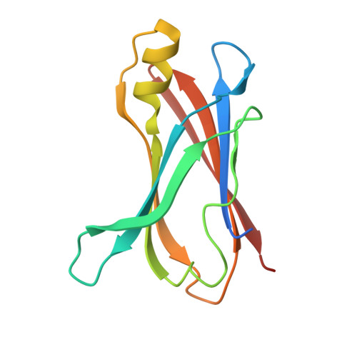
Role of the glutamic acid 54 residue in transthyretin stability and thyroxine binding
Miyata, M., Sato, T., Mizuguchi, M., Nakamura, T., Ikemizu, S., Nabeshima, Y., Susuki, S., Suwa, Y., Morioka, H., Ando, Y., Suico, M.A., Shuto, T., Koga, T., Yamagata, Y., Kai, H.(2010) Biochemistry 49: 114-123
- PubMed: 19950966
- DOI: https://doi.org/10.1021/bi901677z
- Primary Citation of Related Structures:
3A4D, 3A4E, 3A4F - PubMed Abstract:
Transthyretin (TTR) is a tetrameric protein associated with amyloidosis caused by tetramer dissociation and monomer misfolding. The structure of two TTR variants (E54G and E54K) with Glu54 point mutation that cause clinically aggressive amyloidosis remains unclear, although amyloidogenicity of artificial triple mutations (residues 53-55) in beta-strand D had been investigated. Here we first analyzed the crystal structures and biochemical and biophysical properties of E54G and E54K TTRs. The direction of the Lys15 side chain in E54K TTR and the surface electrostatic potential in the edge region in both variants were different from those of wild-type TTR. The presence of Lys54 leads to destabilization of tetramer structure due to enhanced electrostatic repulsion between Lys15 of two monomers. Consistent with structural data, the biochemical analyses demonstrated that E54G and E54K TTRs were more unstable than wild-type TTR. Furthermore, the entrance of the thyroxine (T(4)) binding pocket in TTR was markedly narrower in E54K TTR and wider in E54G TTR compared with wild-type TTR. The tetramer stabilization and amyloid fibril formation assays in the presence of T(4) showed lower tetramer stability and more fibril formation in E54K and E54G TTRs than in wild-type TTR, suggesting decreased T(4) binding to the TTR variants. These findings indicate that structural modification by Glu54 point mutation may sufficiently alter tetramer stability and T(4) binding.
Organizational Affiliation:
Department of Molecular Medicine, Global COE Cell Fate Regulation Research and Education Unit, Kumamoto University, 5-1 Oe-Honmachi, Kumamoto 862-0973, Japan.


