3D1J

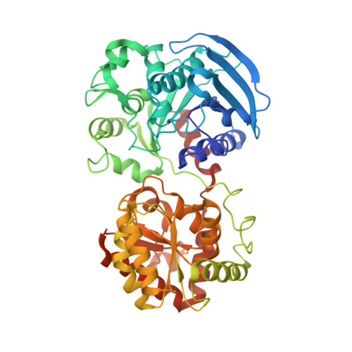
Crystal Structure of E.coli GS mutant dmGS(C7S;C408S)


Experimental Data Snapshot

  • Method: X-RAY DIFFRACTION
  • Resolution: 3.00 Å
  • R-Value Free: 0.263 
  • R-Value Work: 0.209 
  • R-Value Observed: 0.212 

Starting Model: experimental
View more details

wwPDB Validation   3D Report Full Report


This is version 1.3 of the entry. See complete history


Literature

The Crystal Structures of the Open and Catalytically Competent Closed Conformation of Escherichia coli Glycogen Synthase.

Sheng, F.Jia, X.Yep, A.Preiss, J.Geiger, J.H.

(2009) J Biol Chem 284: 17796-17807

  • DOI: https://doi.org/10.1074/jbc.M809804200
  • Primary Citation of Related Structures:  
    2QZS, 2R4T, 2R4U, 3COP, 3D1J, 3GUH

  • PubMed Abstract: 

    Escherichia coli glycogen synthase (EcGS, EC 2.4.1.21) is a retaining glycosyltransferase (GT) that transfers glucose from adenosine diphosphate glucose to a glucan chain acceptor with retention of configuration at the anomeric carbon. EcGS belongs to the GT-B structural superfamily. Here we report several EcGS x-ray structures that together shed considerable light on the structure and function of these enzymes. The structure of the wild-type enzyme bound to ADP and glucose revealed a 15.2 degrees overall domain-domain closure and provided for the first time the structure of the catalytically active, closed conformation of a glycogen synthase. The main chain carbonyl group of His-161, Arg-300, and Lys-305 are suggested by the structure to act as critical catalytic residues in the transglycosylation. Glu-377, previously thought to be catalytic is found on the alpha-face of the glucose and plays an electrostatic role in the active site and as a glucose ring locator. This is also consistent with the structure of the EcGS(E377A)-ADP-HEPPSO complex where the glucose moiety is either absent or disordered in the active site.


  • Organizational Affiliation

    Department of Chemistry, Michigan State University, East Lansing, MI 48824, USA.


Macromolecules
Find similar proteins by:  (by identity cutoff)  |  3D Structure
Entity ID: 1
MoleculeChains Sequence LengthOrganismDetailsImage
Glycogen synthase477Escherichia coliMutation(s): 2 
Gene Names: glgA
EC: 2.4.1.21
UniProt
Find proteins for P0A6U8 (Escherichia coli (strain K12))
Explore P0A6U8 
Go to UniProtKB:  P0A6U8
Entity Groups  
Sequence Clusters30% Identity50% Identity70% Identity90% Identity95% Identity100% Identity
UniProt GroupP0A6U8
Sequence Annotations
Expand
  • Reference Sequence
Experimental Data & Validation

Experimental Data

  • Method: X-RAY DIFFRACTION
  • Resolution: 3.00 Å
  • R-Value Free: 0.263 
  • R-Value Work: 0.209 
  • R-Value Observed: 0.212 
  • Space Group: H 3 2
Unit Cell:
Length ( Å )Angle ( ˚ )
a = 232.959α = 90
b = 232.959β = 90
c = 85.539γ = 120
Software Package:
Software NamePurpose
MOLREPphasing
REFMACrefinement
PDB_EXTRACTdata extraction
HKL-2000data collection
HKL-2000data reduction
SCALEPACKdata scaling

Structure Validation

View Full Validation Report


Entry History 

Deposition Data

Revision History  (Full details and data files)

  • Version 1.0: 2009-03-24
    Type: Initial release
  • Version 1.1: 2011-07-13
    Changes: Version format compliance
  • Version 1.2: 2021-10-20
    Changes: Database references
  • Version 1.3: 2023-08-30
    Changes: Data collection, Refinement description