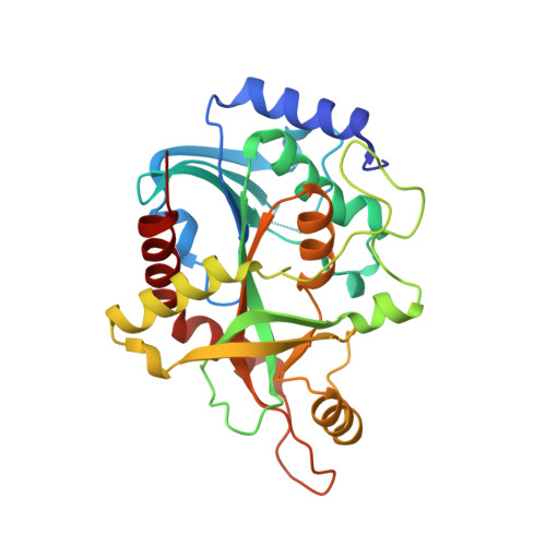
3E0Q

Crystal structure of Schistosoma mansoni purine nucleoside phosphorylase complexed with a novel monocyclic inhibitor


Experimental Data Snapshot

  • Method: X-RAY DIFFRACTION
  • Resolution: 1.90 Å
  • R-Value Free: 0.236 
  • R-Value Work: 0.179 
  • R-Value Observed: 0.182 

Starting Model: experimental
View more details

wwPDB Validation   3D Report Full Report


Ligand Structure Quality Assessment 


This is version 1.2 of the entry. See complete history


Literature

Crystal structure of Schistosoma purine nucleoside phosphorylase complexed with a novel monocyclic inhibitor.

Pereira, H.M.Berdini, V.Ferri, M.R.Cleasby, A.Garratt, R.C.

(2010) Acta Trop 114: 97-102

  • DOI: https://doi.org/10.1016/j.actatropica.2010.01.010
  • Primary Citation of Related Structures:  
    3E0Q

  • PubMed Abstract: 

    A novel inhibitor of Schistosoma PNP was identified using an "in silico" approach allied to enzyme inhibition assays. The compound has a monocyclic structure which has not been previously described for PNP inhibitors. The crystallographic structure of the complex was determined and used to elucidate the binding mode within the active site. Furthermore, the predicted pose was very similar to that determined crystallographically, validating the methodology. The compound Sm_VS1, despite its low molecular weight, possesses an IC(50) of 1.3 microM, surprisingly low when compared with purine analogues. This is presumably due to the formation of eight hydrogen bonds with key residues in the active site E203, N245 and T244. The results of this study highlight the importance of the use of multiple conformations for the target during virtual screening. Indeed the Sm_VS1 compound was only identified after flipping the N245 side chain. It is expected that the structure will be of use in the development of new highly active non-purine based compounds against the Schistosoma enzyme.


  • Organizational Affiliation

    Instituto de Física de São Carlos, Universidade de São Paulo, Brazil. hmuniz.pereira@gmail.com


Macromolecules
Find similar proteins by:  (by identity cutoff)  |  3D Structure
Entity ID: 1
MoleculeChains Sequence LengthOrganismDetailsImage
Purine-nucleoside phosphorylase
A, B, C
287Schistosoma mansoniMutation(s): 0 
Gene Names: SmPNP
EC: 2.4.2.1
UniProt
Find proteins for Q9BMI9 (Schistosoma mansoni)
Explore Q9BMI9 
Go to UniProtKB:  Q9BMI9
Entity Groups  
Sequence Clusters30% Identity50% Identity70% Identity90% Identity95% Identity100% Identity
UniProt GroupQ9BMI9
Sequence Annotations
Expand
  • Reference Sequence
Experimental Data & Validation

Experimental Data

  • Method: X-RAY DIFFRACTION
  • Resolution: 1.90 Å
  • R-Value Free: 0.236 
  • R-Value Work: 0.179 
  • R-Value Observed: 0.182 
  • Space Group: P 21 21 21
Unit Cell:
Length ( Å )Angle ( ˚ )
a = 47.849α = 90
b = 120.018β = 90
c = 131.399γ = 90
Software Package:
Software NamePurpose
MOSFLMdata reduction
SCALAdata scaling
PHENIXrefinement
PDB_EXTRACTdata extraction
ADSCdata collection
REFMACphasing

Structure Validation

View Full Validation Report


Ligand Structure Quality Assessment 


Entry History 

Deposition Data

Revision History  (Full details and data files)

  • Version 1.0: 2009-08-18
    Type: Initial release
  • Version 1.1: 2011-07-13
    Changes: Version format compliance
  • Version 1.2: 2023-08-30
    Changes: Data collection, Database references, Derived calculations, Refinement description