3GHS

Human aldose reductase in complex with NADP+ and the inhibitor IDD594. Investigation of global effects of radiation damage on protein structure. Second stage of radiation damage.


Experimental Data Snapshot

  • Method: X-RAY DIFFRACTION
  • Resolution: 1.00 Å
  • R-Value Free: 0.098 
  • R-Value Work: 0.081 

Starting Model: experimental
View more details

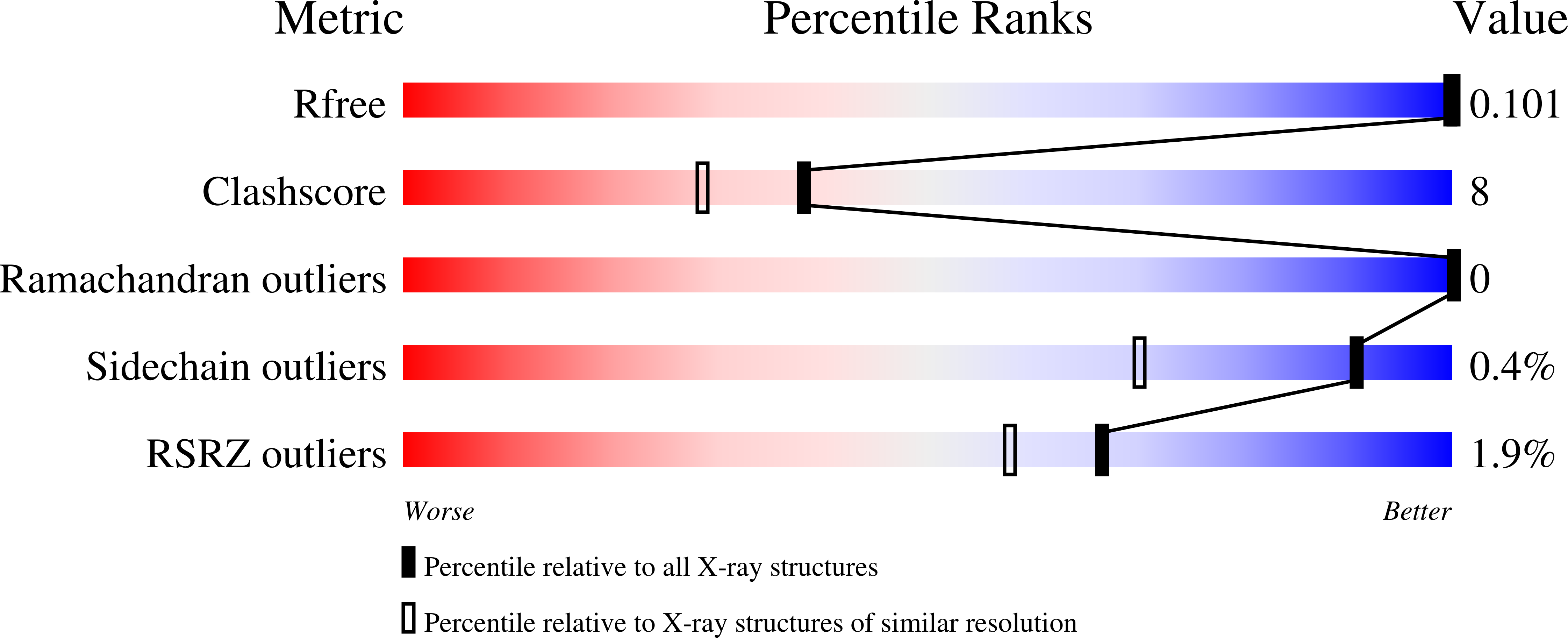
wwPDB Validation   3D Report Full Report


Ligand Structure Quality Assessment 


This is version 1.2 of the entry. See complete history


Literature

X-ray-radiation-induced cooperative atomic movements in protein.

Petrova, T.Lunin, V.Y.Ginell, S.Hazemann, I.Lazarski, K.Mitschler, A.Podjarny, A.Joachimiak, A.

(2009) J Mol Biol 387: 1092-1105

  • DOI: https://doi.org/10.1016/j.jmb.2009.02.030
  • Primary Citation of Related Structures:  
    3GHR, 3GHS, 3GHT, 3GHU

  • PubMed Abstract: 

    X-rays interact with biological matter and cause damage. Proteins and other macromolecules are damaged primarily by ionizing X-ray photons and secondarily by reactive radiolytic chemical species. In particular, protein molecules are damaged during X-ray diffraction experiments with protein crystals, which is, in many cases, a serious hindrance to structure solution. The local X-ray-induced structural changes of the protein molecule have been studied using a number of model systems. However, it is still not well understood whether these local chemical changes lead to global structural changes in protein and what the mechanism is. We present experimental evidence at atomic resolution indicating the movement of large parts of the protein globule together with bound water molecules in the early stages of radiation damage to the protein crystal. The data were obtained from a crystal cryocooled to approximately 100 K and diffracting to 1 A. The movement of the protein structural elements occurs simultaneously with the decarboxylation of several glutamate and aspartate residues that mediate contacts between moving protein structural elements and with the rearrangement of the water network. The analysis of the anisotropy of atomic displacement parameters reveals that the observed atomic movements occur at different rates in different unit cells of the crystal. Thus, the examination of the cooperative atomic movement enables us to better understand how radiation-induced local chemical and structural changes of the protein molecule eventually lead to disorder in protein crystals.


  • Organizational Affiliation

    Structural Biology Center, Biosciences Division, Argonne National Laboratory, Argonne, IL 60439, USA.


Macromolecules
Find similar proteins by:  (by identity cutoff)  |  3D Structure
Entity ID: 1
MoleculeChains Sequence LengthOrganismDetailsImage
Aldose reductase316Homo sapiensMutation(s): 0 
Gene Names: AKR1B1ALDR1
EC: 1.1.1.21 (PDB Primary Data), 1.1.1.372 (UniProt), 1.1.1.300 (UniProt), 1.1.1.54 (UniProt)
UniProt & NIH Common Fund Data Resources
Find proteins for P15121 (Homo sapiens)
Explore P15121 
Go to UniProtKB:  P15121
PHAROS:  P15121
GTEx:  ENSG00000085662 
Entity Groups  
Sequence Clusters30% Identity50% Identity70% Identity90% Identity95% Identity100% Identity
UniProt GroupP15121
Sequence Annotations
Expand
  • Reference Sequence
Small Molecules
Binding Affinity Annotations 
IDSourceBinding Affinity
LDT BindingDB:  3GHS IC50: min: 3, max: 30 (nM) from 2 assay(s)
-TΔS: min: -2.26e+1, max: 7.99 (kJ/mol) from 3 assay(s)
ΔH: -4.62e+1 (kJ/mol) from 1 assay(s)
ΔG: min: -4.09e+1, max: -3.13e+1 (kJ/mol) from 4 assay(s)
Experimental Data & Validation

Experimental Data

  • Method: X-RAY DIFFRACTION
  • Resolution: 1.00 Å
  • R-Value Free: 0.098 
  • R-Value Work: 0.081 
  • Space Group: P 1 21 1
Unit Cell:
Length ( Å )Angle ( ˚ )
a = 49.368α = 90
b = 66.801β = 92.22
c = 47.355γ = 90
Software Package:
Software NamePurpose
HKL-3000data collection
AMoREphasing
SHELXL-97refinement
HKL-3000data reduction
HKL-3000data scaling

Structure Validation

View Full Validation Report


Ligand Structure Quality Assessment 


Entry History 

Deposition Data

Revision History  (Full details and data files)

  • Version 1.0: 2009-03-24
    Type: Initial release
  • Version 1.1: 2011-07-13
    Changes: Version format compliance
  • Version 1.2: 2023-09-06
    Changes: Data collection, Database references, Derived calculations, Refinement description