3MOR

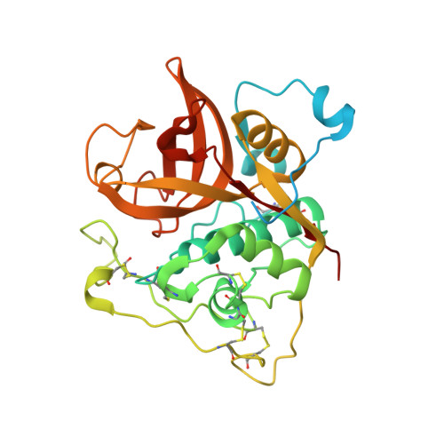
Crystal structure of Cathepsin B from Trypanosoma Brucei


Experimental Data Snapshot

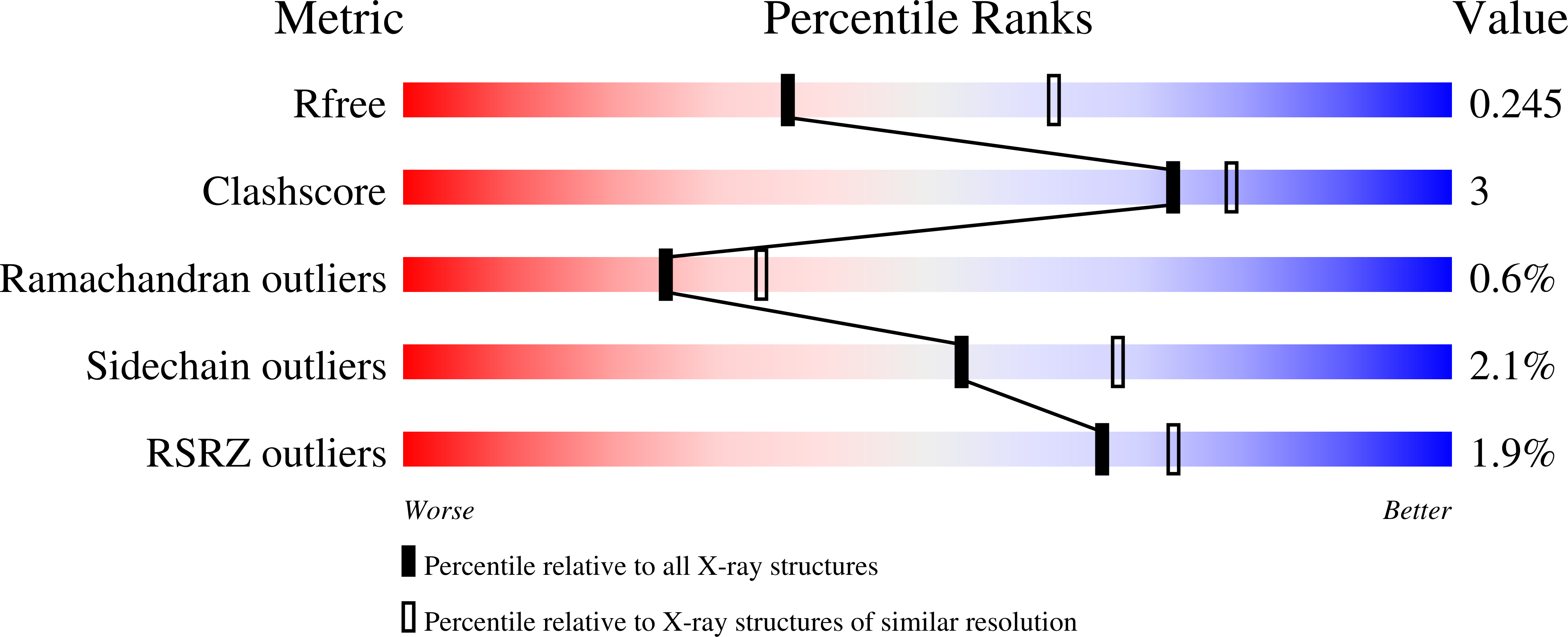
  • Method: X-RAY DIFFRACTION
  • Resolution: 2.55 Å
  • R-Value Free: 0.243 
  • R-Value Work: 0.202 
  • R-Value Observed: 0.204 

Starting Model: experimental
View more details

wwPDB Validation   3D Report Full Report


This is version 2.2 of the entry. See complete history


Literature

In vivo protein crystallization opens new routes in structural biology.

Koopmann, R.Cupelli, K.Redecke, L.Nass, K.Deponte, D.P.White, T.A.Stellato, F.Rehders, D.Liang, M.Andreasson, J.Aquila, A.Bajt, S.Barthelmess, M.Barty, A.Bogan, M.J.Bostedt, C.Boutet, S.Bozek, J.D.Caleman, C.Coppola, N.Davidsson, J.Doak, R.B.Ekeberg, T.Epp, S.W.Erk, B.Fleckenstein, H.Foucar, L.Graafsma, H.Gumprecht, L.Hajdu, J.Hampton, C.Y.Hartmann, A.Hartmann, R.Hauser, G.Hirsemann, H.Holl, P.Hunter, M.S.Kassemeyer, S.Kirian, R.A.Lomb, L.Maia, F.R.Kimmel, N.Martin, A.V.Messerschmidt, M.Reich, C.Rolles, D.Rudek, B.Rudenko, A.Schlichting, I.Schulz, J.Seibert, M.M.Shoeman, R.L.Sierra, R.G.Soltau, H.Stern, S.Struder, L.Timneanu, N.Ullrich, J.Wang, X.Weidenspointner, G.Weierstall, U.Williams, G.J.Wunderer, C.B.Fromme, P.Spence, J.C.Stehle, T.Chapman, H.N.Betzel, C.Duszenko, M.

(2012) Nat Methods 9: 259-262

  • DOI: https://doi.org/10.1038/nmeth.1859
  • Primary Citation of Related Structures:  
    3MOR

  • PubMed Abstract: 

    Protein crystallization in cells has been observed several times in nature. However, owing to their small size these crystals have not yet been used for X-ray crystallographic analysis. We prepared nano-sized in vivo-grown crystals of Trypanosoma brucei enzymes and applied the emerging method of free-electron laser-based serial femtosecond crystallography to record interpretable diffraction data. This combined approach will open new opportunities in structural systems biology.


  • Organizational Affiliation

    Interfaculty Institute of Biochemistry, University of Tübingen, Tübingen, Germany.


Macromolecules
Find similar proteins by:  (by identity cutoff)  |  3D Structure
Entity ID: 1
MoleculeChains Sequence LengthOrganismDetailsImage
Cathepsin B-like cysteine protease
A, B
317Trypanosoma bruceiMutation(s): 0 
Gene Names: Tb927.6.560
EC: 3.4.22
UniProt
Find proteins for Q6R7Z5 (Trypanosoma brucei)
Explore Q6R7Z5 
Go to UniProtKB:  Q6R7Z5
Entity Groups  
Sequence Clusters30% Identity50% Identity70% Identity90% Identity95% Identity100% Identity
UniProt GroupQ6R7Z5
Glycosylation
Glycosylation Sites: 1
Sequence Annotations
Expand
  • Reference Sequence
Oligosaccharides

Help

Entity ID: 2
MoleculeChains Length2D Diagram Glycosylation3D Interactions
2-acetamido-2-deoxy-beta-D-glucopyranose-(1-4)-2-acetamido-2-deoxy-beta-D-glucopyranose
C, D
2N-Glycosylation
Glycosylation Resources
GlyTouCan:  G42666HT
GlyCosmos:  G42666HT
GlyGen:  G42666HT
Small Molecules
Ligands 3 Unique
IDChains Name / Formula / InChI Key2D Diagram3D Interactions
GOL
Query on GOL

Download Ideal Coordinates CCD File 
E [auth A]
F [auth A]
H [auth A]
I [auth B]
J [auth B]
E [auth A],
F [auth A],
H [auth A],
I [auth B],
J [auth B],
K [auth B]
GLYCEROL
C3 H8 O3
PEDCQBHIVMGVHV-UHFFFAOYSA-N
CL
Query on CL

Download Ideal Coordinates CCD File 
M [auth B]CHLORIDE ION
Cl
VEXZGXHMUGYJMC-UHFFFAOYSA-M
NA
Query on NA

Download Ideal Coordinates CCD File 
G [auth A],
L [auth B]
SODIUM ION
Na
FKNQFGJONOIPTF-UHFFFAOYSA-N
Experimental Data & Validation

Experimental Data

  • Method: X-RAY DIFFRACTION
  • Resolution: 2.55 Å
  • R-Value Free: 0.243 
  • R-Value Work: 0.202 
  • R-Value Observed: 0.204 
  • Space Group: P 1 21 1
Unit Cell:
Length ( Å )Angle ( ˚ )
a = 53.91α = 90
b = 75.49β = 104.8
c = 75.6γ = 90
Software Package:
Software NamePurpose
REFMACrefinement
PDB_EXTRACTdata extraction
XDSdata scaling
XDSdata reduction
PHASERphasing

Structure Validation

View Full Validation Report


Entry History 

Deposition Data

Revision History  (Full details and data files)

  • Version 1.0: 2011-11-02
    Type: Initial release
  • Version 1.1: 2012-06-20
    Changes: Database references
  • Version 2.0: 2020-07-29
    Type: Remediation
    Reason: Carbohydrate remediation
    Changes: Atomic model, Data collection, Derived calculations, Structure summary
  • Version 2.1: 2023-09-06
    Changes: Data collection, Database references, Refinement description, Structure summary
  • Version 2.2: 2024-11-06
    Changes: Structure summary