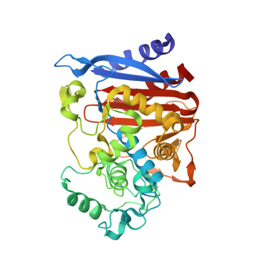
3O87

Crystal structure of AmpC beta-lactamase in complex with a sulfonamide boronic acid inhibitor


Experimental Data Snapshot

  • Method: X-RAY DIFFRACTION
  • Resolution: 1.78 Å
  • R-Value Free: 0.232 
  • R-Value Work: 0.188 
  • R-Value Observed: 0.189 

Starting Model: experimental
View more details

wwPDB Validation   3D Report Full Report


Ligand Structure Quality Assessment 


This is version 1.3 of the entry. See complete history


Literature

Design, Synthesis, Crystal Structures, and Antimicrobial Activity of Sulfonamide Boronic Acids as beta-Lactamase Inhibitors

Eidam, O.Romagnoli, C.Caselli, E.Babaoglu, K.Pohlhaus, D.T.Karpiak, J.Bonnet, R.Shoichet, B.K.Prati, F.

(2010) J Med Chem 53: 7852-7863

  • DOI: https://doi.org/10.1021/jm101015z
  • Primary Citation of Related Structures:  
    3O86, 3O87, 3O88

  • PubMed Abstract: 

    We investigated a series of sulfonamide boronic acids that resulted from the merging of two unrelated AmpC β-lactamase inhibitor series. The new boronic acids differed in the replacement of the canonical carboxamide, found in all penicillin and cephalosporin antibiotics, with a sulfonamide. Surprisingly, these sulfonamides had a highly distinct structure-activity relationship from the previously explored carboxamides, high ligand efficiencies (up to 0.91), and K(i) values down to 25 nM and up to 23 times better for smaller analogues. Conversely, K(i) values were 10-20 times worse for larger molecules than in the carboxamide congener series. X-ray crystal structures (1.6-1.8 Å) of AmpC with three of the new sulfonamides suggest that this altered structure-activity relationship results from the different geometry and polarity of the sulfonamide versus the carboxamide. The most potent inhibitor reversed β-lactamase-mediated resistance to third generation cephalosporins, lowering their minimum inhibitory concentrations up to 32-fold in cell culture.


  • Organizational Affiliation

    Department of Pharmaceutical Chemistry, Byers Hall, University of California San Francisco, 1700 4th Street, San Francisco, California 94158, United States.


Macromolecules
Find similar proteins by:  (by identity cutoff)  |  3D Structure
Entity ID: 1
MoleculeChains Sequence LengthOrganismDetailsImage
Beta-lactamase
A, B
358Escherichia coli K-12Mutation(s): 0 
Gene Names: ampAampCb4150JW4111
EC: 3.5.2.6
UniProt
Find proteins for P00811 (Escherichia coli (strain K12))
Explore P00811 
Go to UniProtKB:  P00811
Entity Groups  
Sequence Clusters30% Identity50% Identity70% Identity90% Identity95% Identity100% Identity
UniProt GroupP00811
Sequence Annotations
Expand
  • Reference Sequence
Small Molecules
Binding Affinity Annotations 
IDSourceBinding Affinity
BSG PDBBind:  3O87 Ki: 25 (nM) from 1 assay(s)
Experimental Data & Validation

Experimental Data

  • Method: X-RAY DIFFRACTION
  • Resolution: 1.78 Å
  • R-Value Free: 0.232 
  • R-Value Work: 0.188 
  • R-Value Observed: 0.189 
  • Space Group: C 1 2 1
Unit Cell:
Length ( Å )Angle ( ˚ )
a = 118.13α = 90
b = 76.19β = 116.16
c = 97.38γ = 90
Software Package:
Software NamePurpose
XSCALEdata processing
PHENIXrefinement
PDB_EXTRACTdata extraction
Blu-Icedata collection
XDSdata reduction
XSCALEdata scaling
PHENIXphasing

Structure Validation

View Full Validation Report


Ligand Structure Quality Assessment 


Entry History 

Deposition Data

Revision History  (Full details and data files)

  • Version 1.0: 2010-11-03
    Type: Initial release
  • Version 1.1: 2011-07-13
    Changes: Version format compliance
  • Version 1.2: 2013-06-19
    Changes: Database references
  • Version 1.3: 2023-09-06
    Changes: Data collection, Database references, Derived calculations, Refinement description