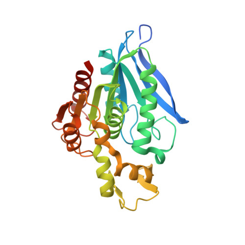
3PFB

Crystal structure of the Lactobacillus johnsonii cinnamoyl esterase LJ0536 S106A mutant in complex with ethylferulate


Experimental Data Snapshot

  • Method: X-RAY DIFFRACTION
  • Resolution: 1.58 Å
  • R-Value Free: 0.304 
  • R-Value Work: 0.211 
  • R-Value Observed: 0.216 

Starting Model: experimental
View more details

wwPDB Validation   3D Report Full Report


Ligand Structure Quality Assessment 


This is version 1.3 of the entry. See complete history


Literature

An Inserted alpha/beta Subdomain Shapes the Catalytic Pocket of Lactobacillus johnsonii Cinnamoyl Esterase

Lai, K.K.Stogios, P.J.Vu, C.Xu, X.Cui, H.Molloy, S.Savchenko, A.Yakunin, A.Gonzalez, C.F.

(2011) PLoS One 6: e23269-e23269

  • DOI: https://doi.org/10.1371/journal.pone.0023269
  • Primary Citation of Related Structures:  
    3PF8, 3PF9, 3PFB, 3PFC, 3QM1, 3S2Z

  • PubMed Abstract: 

    Microbial enzymes produced in the gastrointestinal tract are primarily responsible for the release and biochemical transformation of absorbable bioactive monophenols. In the present work we described the crystal structure of LJ0536, a serine cinnamoyl esterase produced by the probiotic bacterium Lactobacillus johnsonii N6.2. We crystallized LJ0536 in the apo form and in three substrate-bound complexes. The structure showed a canonical α/β fold characteristic of esterases, and the enzyme is dimeric. Two classical serine esterase motifs (GlyXSerXGly) can be recognized from the amino acid sequence, and the structure revealed that the catalytic triad of the enzyme is formed by Ser(106), His(225), and Asp(197), while the other motif is non-functional. In all substrate-bound complexes, the aromatic acyl group of the ester compound was bound in the deepest part of the catalytic pocket. The binding pocket also contained an unoccupied area that could accommodate larger ligands. The structure revealed a prominent inserted α/β subdomain of 54 amino acids, from which multiple contacts to the aromatic acyl groups of the substrates are made. Inserts of this size are seen in other esterases, but the secondary structure topology of this subdomain of LJ0536 is unique to this enzyme and its closest homolog (Est1E) in the Protein Databank. The binding mechanism characterized (involving the inserted α/β subdomain) clearly differentiates LJ0536 from enzymes with similar activity of a fungal origin. The structural features herein described together with the activity profile of LJ0536 suggest that this enzyme should be clustered in a new group of bacterial cinnamoyl esterases.


  • Organizational Affiliation

    Department of Microbiology and Cell Science, Genetics Institute, University of Florida, Gainesville, Florida, United States of America.


Macromolecules
Find similar proteins by:  (by identity cutoff)  |  3D Structure
Entity ID: 1
MoleculeChains Sequence LengthOrganismDetailsImage
Cinnamoyl esterase
A, B
270Lactobacillus johnsoniiMutation(s): 1 
Gene Names: LJ0536
EC: 3.1.1
UniProt
Find proteins for D3YEX6 (Lactobacillus johnsonii)
Explore D3YEX6 
Go to UniProtKB:  D3YEX6
Entity Groups  
Sequence Clusters30% Identity50% Identity70% Identity90% Identity95% Identity100% Identity
UniProt GroupD3YEX6
Sequence Annotations
Expand
  • Reference Sequence
Small Molecules
Ligands 3 Unique
IDChains Name / Formula / InChI Key2D Diagram3D Interactions
ZYC
Query on ZYC

Download Ideal Coordinates CCD File 
I [auth A],
X [auth B]
ethyl (2E)-3-(4-hydroxy-3-methoxyphenyl)prop-2-enoate
C12 H14 O4
ATJVZXXHKSYELS-FNORWQNLSA-N
CL
Query on CL

Download Ideal Coordinates CCD File 
H [auth A],
U [auth B],
V [auth B],
W [auth B]
CHLORIDE ION
Cl
VEXZGXHMUGYJMC-UHFFFAOYSA-M
NH4
Query on NH4

Download Ideal Coordinates CCD File 
C [auth A]
D [auth A]
E [auth A]
F [auth A]
G [auth A]
C [auth A],
D [auth A],
E [auth A],
F [auth A],
G [auth A],
J [auth B],
K [auth B],
L [auth B],
M [auth B],
N [auth B],
O [auth B],
P [auth B],
Q [auth B],
R [auth B],
S [auth B],
T [auth B]
AMMONIUM ION
H4 N
QGZKDVFQNNGYKY-UHFFFAOYSA-O
Experimental Data & Validation

Experimental Data

  • Method: X-RAY DIFFRACTION
  • Resolution: 1.58 Å
  • R-Value Free: 0.304 
  • R-Value Work: 0.211 
  • R-Value Observed: 0.216 
  • Space Group: C 1 2 1
Unit Cell:
Length ( Å )Angle ( ˚ )
a = 72.26α = 90
b = 83.891β = 98.21
c = 88.947γ = 90
Software Package:
Software NamePurpose
CrystalCleardata collection
PHASERphasing
REFMACrefinement
HKL-2000data reduction
HKL-2000data scaling

Structure Validation

View Full Validation Report


Ligand Structure Quality Assessment 


Entry History 

Deposition Data

Revision History  (Full details and data files)

  • Version 1.0: 2011-08-31
    Type: Initial release
  • Version 1.1: 2012-10-24
    Changes: Database references
  • Version 1.2: 2024-02-21
    Changes: Data collection, Database references, Derived calculations
  • Version 1.3: 2024-04-03
    Changes: Refinement description