3RK0 | pdb_00003rk0

X-ray crystal Structure of the putative N-type ATP pyrophosphatase (PF0828) in complex with AMP from Pyrococcus furiosus, Northeast Structural Genomics Consortium Target PfR23


Experimental Data Snapshot

  • Method: X-RAY DIFFRACTION
  • Resolution: 2.40 Å
  • R-Value Free: 
    0.278 (Depositor), 0.290 (DCC) 
  • R-Value Work: 
    0.229 (Depositor), 0.240 (DCC) 
  • R-Value Observed: 
    0.229 (Depositor) 

Starting Model: experimental
View more details

wwPDB Validation   3D Report Full Report


Ligand Structure Quality Assessment 

Created with Raphaël 2.3.0Worse 01 BetterLigand structure goodness of fit to experimental dataBest fitted AMPClick on this verticalbar to view details

This is version 1.5 of the entry. See complete history


Literature

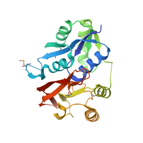
A large conformational change in the putative ATP pyrophosphatase PF0828 induced by ATP binding.

Forouhar, F.Saadat, N.Hussain, M.Seetharaman, J.Lee, I.Janjua, H.Xiao, R.Shastry, R.Acton, T.B.Montelione, G.T.Tong, L.

(2011) Acta Crystallogr Sect F Struct Biol Cryst Commun 67: 1323-1327

  • DOI: https://doi.org/10.1107/S1744309111031447
  • Primary Citation of Related Structures:  
    3RJZ, 3RK0, 3RK1

  • PubMed Abstract: 

    ATP pyrophosphatases (ATP PPases) are widely distributed in archaea and eukaryotes. They share an HUP domain at the N-terminus with a conserved PP-motif that interacts with the phosphates of ATP. The PF0828 protein from Pyrococcus furiosus is a member of the ATP PPase superfamily and it also has a 100-residue C-terminal extension that contains a strictly conserved EGG(E/D)xE(T/S) motif, which has been named the EGT-motif. Here, crystal structures of PF0828 alone and in complex with ATP or AMP are reported. The HUP domain contains a central five-stranded β-sheet that is surrounded by four helices, as in other related structures. The C-terminal extension forms a separate domain, named the EGT domain, which makes tight interactions with the HUP domain, bringing the EGT-motif near to the PP-motif and defining the putative active site of PF0828. Both motifs interact with the phosphate groups of ATP. A loop in the HUP domain undergoes a large conformational change to recognize the adenine base of ATP. In solution and in the crystal PF0828 is a dimer formed by the side-by-side arrangement of the HUP domains of the two monomers. The putative active site is located far from the dimer interface.


  • Organizational Affiliation

    Northeast Structural Genomics Consortium, USA.


Macromolecules
Find similar proteins by:  (by identity cutoff)  |  3D Structure
Entity ID: 1
MoleculeChains Sequence LengthOrganismDetailsImage
N-type ATP pyrophosphatase superfamily237Pyrococcus furiosus DSM 3638Mutation(s): 0 
Gene Names: PF0828
UniProt
Find proteins for Q8U2K6 (Pyrococcus furiosus (strain ATCC 43587 / DSM 3638 / JCM 8422 / Vc1))
Explore Q8U2K6 
Go to UniProtKB:  Q8U2K6
Entity Groups  
Sequence Clusters30% Identity50% Identity70% Identity90% Identity95% Identity100% Identity
UniProt GroupQ8U2K6
Sequence Annotations
Expand
  • Reference Sequence
Small Molecules
Ligands 1 Unique
IDChains Name / Formula / InChI Key2D Diagram3D Interactions
AMP
Query on AMP

Download Ideal Coordinates CCD File 
B [auth A]ADENOSINE MONOPHOSPHATE
C10 H14 N5 O7 P
UDMBCSSLTHHNCD-KQYNXXCUSA-N
Modified Residues  1 Unique
IDChains TypeFormula2D DiagramParent
MSE
Query on MSE
A
L-PEPTIDE LINKINGC5 H11 N O2 SeMET
Experimental Data & Validation

Experimental Data

  • Method: X-RAY DIFFRACTION
  • Resolution: 2.40 Å
  • R-Value Free:  0.278 (Depositor), 0.290 (DCC) 
  • R-Value Work:  0.229 (Depositor), 0.240 (DCC) 
  • R-Value Observed: 0.229 (Depositor) 
Space Group: P 41 21 2
Unit Cell:
Length ( Å )Angle ( ˚ )
a = 85.201α = 90
b = 85.201β = 90
c = 74.249γ = 90
Software Package:
Software NamePurpose
ADSCdata collection
COMOphasing
CNSrefinement
HKL-2000data reduction
SCALEPACKdata scaling

Structure Validation

View Full Validation Report


Ligand Structure Quality Assessment 

Created with Raphaël 2.3.0Worse 01 BetterLigand structure goodness of fit to experimental dataBest fitted AMPClick on this verticalbar to view details

Entry History 

Revision History  (Full details and data files)

  • Version 1.0: 2011-05-11
    Type: Initial release
  • Version 1.1: 2011-07-13
    Changes: Version format compliance
  • Version 1.2: 2012-01-11
    Changes: Database references
  • Version 1.3: 2023-09-13
    Changes: Data collection, Database references, Derived calculations, Refinement description
  • Version 1.4: 2023-12-06
    Changes: Data collection
  • Version 1.5: 2024-10-30
    Changes: Structure summary