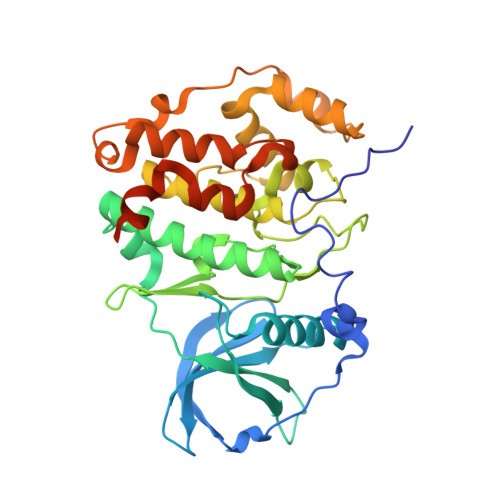
3U87

Structure of a chimeric construct of human CK2alpha and human CK2alpha' in complex with a non-hydrolysable ATP-analogue


Experimental Data Snapshot

  • Method: X-RAY DIFFRACTION
  • Resolution: 2.90 Å
  • R-Value Free: 0.218 
  • R-Value Work: 0.186 
  • R-Value Observed: 0.187 

Starting Model: experimental
View more details

wwPDB Validation   3D Report Full Report


Ligand Structure Quality Assessment 


This is version 1.3 of the entry. See complete history


Literature

Low-density crystal packing of human protein kinase CK2 catalytic subunit in complex with resorufin or other ligands: a tool to study the unique hinge-region plasticity of the enzyme without packing bias.

Klopffleisch, K.Issinger, O.G.Niefind, K.

(2012) Acta Crystallogr D Biol Crystallogr 68: 883-892

  • DOI: https://doi.org/10.1107/S0907444912016587
  • Primary Citation of Related Structures:  
    3U87, 3U9C

  • PubMed Abstract: 

    A low-resolution structure of the catalytic subunit CK2α of human protein kinase CK2 (formerly known as casein kinase 2) in complex with the ATP-competitive inhibitor resorufin is presented. The structure supplements previous human CK2α structures in which the interdomain hinge/helix αD region adopts a closed conformation correlating to a canonically established catalytic spine as is typical for eukaryotic protein kinases. In the corresponding crystal packing the hinge/helix αD region is nearly unaffected by crystal contacts, so that largely unbiased conformational adaptions are possible. This is documented by published human CK2α structures with the same crystal packing but with an open hinge/helix αD region, one of which has been redetermined here with a higher symmetry. An overview of all published human CK2α crystal packings serves as the basis for a discussion of the factors that determine whether the open or the closed hinge/helix αD conformation is adopted. Lyotropic salts in crystallization support the closed conformation, in which the Phe121 side chain complements the hydrophobic catalytic spine ensemble. Consequently, genuine ligand effects on the hinge/helix αD conformation can be best studied under moderate salt conditions. Ligands that stabilize either the open or the closed conformation by hydrogen bonds are known, but a general rule is not yet apparent.


  • Organizational Affiliation

    Department für Chemie, Institut für Biochemie, Universität zu Köln, Otto-Fischer-Strasse 12-14, D-50674 Köln, Germany.


Macromolecules
Find similar proteins by:  (by identity cutoff)  |  3D Structure
Entity ID: 1
MoleculeChains Sequence LengthOrganismDetailsImage
Casein kinase II subunit alpha
A, B
349Homo sapiensMutation(s): 0 
Gene Names: CSNK2A1CK2A1
EC: 2.7.11.1
UniProt & NIH Common Fund Data Resources
Find proteins for P68400 (Homo sapiens)
Explore P68400 
Go to UniProtKB:  P68400
PHAROS:  P68400
GTEx:  ENSG00000101266 
Find proteins for P19784 (Homo sapiens)
Explore P19784 
Go to UniProtKB:  P19784
PHAROS:  P19784
GTEx:  ENSG00000070770 
Entity Groups  
Sequence Clusters30% Identity50% Identity70% Identity90% Identity95% Identity100% Identity
UniProt GroupsP68400P19784
Sequence Annotations
Expand
  • Reference Sequence
Small Molecules
Ligands 5 Unique
IDChains Name / Formula / InChI Key2D Diagram3D Interactions
ANP
Query on ANP

Download Ideal Coordinates CCD File 
C [auth A],
I [auth B]
PHOSPHOAMINOPHOSPHONIC ACID-ADENYLATE ESTER
C10 H17 N6 O12 P3
PVKSNHVPLWYQGJ-KQYNXXCUSA-N
SO4
Query on SO4

Download Ideal Coordinates CCD File 
G [auth A],
M [auth B]
SULFATE ION
O4 S
QAOWNCQODCNURD-UHFFFAOYSA-L
GOL
Query on GOL

Download Ideal Coordinates CCD File 
H [auth A],
N [auth B]
GLYCEROL
C3 H8 O3
PEDCQBHIVMGVHV-UHFFFAOYSA-N
CL
Query on CL

Download Ideal Coordinates CCD File 
F [auth A],
L [auth B]
CHLORIDE ION
Cl
VEXZGXHMUGYJMC-UHFFFAOYSA-M
MG
Query on MG

Download Ideal Coordinates CCD File 
D [auth A],
E [auth A],
J [auth B],
K [auth B]
MAGNESIUM ION
Mg
JLVVSXFLKOJNIY-UHFFFAOYSA-N
Experimental Data & Validation

Experimental Data

  • Method: X-RAY DIFFRACTION
  • Resolution: 2.90 Å
  • R-Value Free: 0.218 
  • R-Value Work: 0.186 
  • R-Value Observed: 0.187 
  • Space Group: P 43 21 2
Unit Cell:
Length ( Å )Angle ( ˚ )
a = 127.821α = 90
b = 127.821β = 90
c = 125.333γ = 90
Software Package:
Software NamePurpose
PHENIXrefinement
XDSdata reduction
XSCALEdata scaling

Structure Validation

View Full Validation Report


Ligand Structure Quality Assessment 


Entry History 

Deposition Data

Revision History  (Full details and data files)

  • Version 1.0: 2012-05-30
    Type: Initial release
  • Version 1.1: 2013-01-23
    Changes: Database references
  • Version 1.2: 2017-08-09
    Changes: Source and taxonomy
  • Version 1.3: 2023-09-13
    Changes: Data collection, Database references, Derived calculations, Refinement description