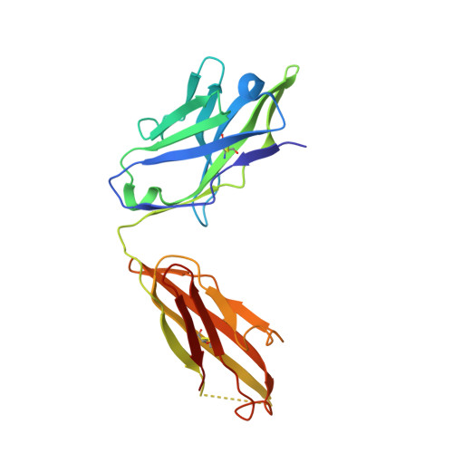
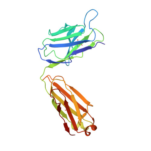
3V4V

crystal structure of a4b7 headpiece complexed with Fab ACT-1 and RO0505376


Experimental Data Snapshot

  • Method: X-RAY DIFFRACTION
  • Resolution: 3.10 Å
  • R-Value Free: 0.231 
  • R-Value Work: 0.197 
  • R-Value Observed: 0.198 

wwPDB Validation   3D Report Full Report


Ligand Structure Quality Assessment 


This is version 2.1 of the entry. See complete history


Literature

Structural specializations of a4b7, an Integrin that Mediates Rolling Adhesion

Yu, Y.Zhu, J.Mi, L.Z.Walz, T.Sun, H.Chen, J.Springer, T.A.

(2012) J Cell Biol 196: 131-146

  • DOI: https://doi.org/10.1083/jcb.201110023
  • Primary Citation of Related Structures:  
    3V4P, 3V4V

  • PubMed Abstract: 

    The lymphocyte homing receptor integrin α(4)β(7) is unusual for its ability to mediate both rolling and firm adhesion. α(4)β(1) and α(4)β(7) are targeted by therapeutics approved for multiple sclerosis and Crohn's disease. Here, we show by electron microscopy and crystallography how two therapeutic Fabs, a small molecule (RO0505376), and mucosal adhesion molecule-1 (MAdCAM-1) bind α(4)β(7). A long binding groove at the α(4)-β(7) interface for immunoglobulin superfamily domains differs in shape from integrin pockets that bind Arg-Gly-Asp motifs. RO0505376 mimics an Ile/Leu-Asp motif in α(4) ligands, and orients differently from Arg-Gly-Asp mimics. A novel auxiliary residue at the metal ion-dependent adhesion site in α(4)β(7) is essential for binding to MAdCAM-1 in Mg(2+) yet swings away when RO0505376 binds. A novel intermediate conformation of the α(4)β(7) headpiece binds MAdCAM-1 and supports rolling adhesion. Lack of induction of the open headpiece conformation by ligand binding enables rolling adhesion to persist until integrin activation is signaled.


  • Organizational Affiliation

    Department of Biological Chemistry and Molecular Pharmacology, Immune Disease Institute and Children's Hospital, Boston, MA 02115, USA.


Macromolecules
Find similar proteins by:  (by identity cutoff)  |  3D Structure
Entity ID: 1
MoleculeChains Sequence LengthOrganismDetailsImage
Integrin alpha-4A,
E [auth C]
597Homo sapiensMutation(s): 1 
Gene Names: ITGA4CD49D
UniProt & NIH Common Fund Data Resources
Find proteins for P13612 (Homo sapiens)
Explore P13612 
Go to UniProtKB:  P13612
PHAROS:  P13612
GTEx:  ENSG00000115232 
Entity Groups  
Sequence Clusters30% Identity50% Identity70% Identity90% Identity95% Identity100% Identity
UniProt GroupP13612
Glycosylation
Glycosylation Sites: 5Go to GlyGen: P13612-1
Sequence Annotations
Expand
  • Reference Sequence
Find similar proteins by:  (by identity cutoff)  |  3D Structure
Entity ID: 2
MoleculeChains Sequence LengthOrganismDetailsImage
Integrin beta-7B,
F [auth D]
503Homo sapiensMutation(s): 0 
Gene Names: ITGB7
UniProt & NIH Common Fund Data Resources
Find proteins for P26010 (Homo sapiens)
Explore P26010 
Go to UniProtKB:  P26010
PHAROS:  P26010
GTEx:  ENSG00000139626 
Entity Groups  
Sequence Clusters30% Identity50% Identity70% Identity90% Identity95% Identity100% Identity
UniProt GroupP26010
Glycosylation
Glycosylation Sites: 2Go to GlyGen: P26010-1
Sequence Annotations
Expand
  • Reference Sequence
Find similar proteins by:  (by identity cutoff)  |  3D Structure
Entity ID: 3
MoleculeChains Sequence LengthOrganismDetailsImage
MONOCLONAL ANTIBODY Act-1 HEAVY CHAINC [auth H],
G [auth M]
219Mus musculusMutation(s): 0 
Entity Groups  
Sequence Clusters30% Identity50% Identity70% Identity90% Identity95% Identity100% Identity
Sequence Annotations
Expand
  • Reference Sequence
Find similar proteins by:  (by identity cutoff)  |  3D Structure
Entity ID: 4
MoleculeChains Sequence LengthOrganismDetailsImage
MONOCLONAL ANTIBODY Act-1 LIGHT CHAIND [auth L],
H [auth N]
217Mus musculusMutation(s): 0 
Entity Groups  
Sequence Clusters30% Identity50% Identity70% Identity90% Identity95% Identity100% Identity
Sequence Annotations
Expand
  • Reference Sequence
Oligosaccharides

Help

Entity ID: 5
MoleculeChains Length2D Diagram Glycosylation3D Interactions
alpha-D-mannopyranose-(1-3)-beta-D-mannopyranose-(1-4)-2-acetamido-2-deoxy-beta-D-glucopyranose-(1-4)-2-acetamido-2-deoxy-beta-D-glucopyranoseI [auth E],
J [auth F]
4N-Glycosylation
Glycosylation Resources
GlyTouCan:  G81315DD
GlyCosmos:  G81315DD
GlyGen:  G81315DD
Small Molecules
Ligands 5 Unique
IDChains Name / Formula / InChI Key2D Diagram3D Interactions
0DU
Query on 0DU

Download Ideal Coordinates CCD File 
JA [auth D],
X [auth B]
N-(2,6-dichlorobenzoyl)-4-[1,6-dimethyl-2-oxo-4-(trifluoromethyl)-1,2-dihydropyridin-3-yl]-L-phenylalanine
C24 H19 Cl2 F3 N2 O4
IBAZQIKVHCLTHH-SFHVURJKSA-N
NAG
Query on NAG

Download Ideal Coordinates CCD File 
BA [auth C]
CA [auth C]
DA [auth C]
EA [auth C]
IA [auth D]
BA [auth C],
CA [auth C],
DA [auth C],
EA [auth C],
IA [auth D],
N [auth A],
O [auth A],
P [auth A],
Q [auth A],
V [auth B],
W [auth B]
2-acetamido-2-deoxy-beta-D-glucopyranose
C8 H15 N O6
OVRNDRQMDRJTHS-FMDGEEDCSA-N
TRS
Query on TRS

Download Ideal Coordinates CCD File 
R [auth A]2-AMINO-2-HYDROXYMETHYL-PROPANE-1,3-DIOL
C4 H12 N O3
LENZDBCJOHFCAS-UHFFFAOYSA-O
CA
Query on CA

Download Ideal Coordinates CCD File 
AA [auth C]
GA [auth D]
HA [auth D]
K [auth A]
L [auth A]
AA [auth C],
GA [auth D],
HA [auth D],
K [auth A],
L [auth A],
M [auth A],
T [auth B],
U [auth B],
Y [auth C],
Z [auth C]
CALCIUM ION
Ca
BHPQYMZQTOCNFJ-UHFFFAOYSA-N
MG
Query on MG

Download Ideal Coordinates CCD File 
FA [auth D],
S [auth B]
MAGNESIUM ION
Mg
JLVVSXFLKOJNIY-UHFFFAOYSA-N
Binding Affinity Annotations 
IDSourceBinding Affinity
0DU PDBBind:  3V4V IC50: 42 (nM) from 1 assay(s)
BindingDB:  3V4V IC50: min: 32, max: 42 (nM) from 2 assay(s)
Experimental Data & Validation

Experimental Data

  • Method: X-RAY DIFFRACTION
  • Resolution: 3.10 Å
  • R-Value Free: 0.231 
  • R-Value Work: 0.197 
  • R-Value Observed: 0.198 
  • Space Group: P 1 21 1
Unit Cell:
Length ( Å )Angle ( ˚ )
a = 127.58α = 90
b = 123.53β = 112.63
c = 154.16γ = 90
Software Package:
Software NamePurpose
HKL-2000data collection
PHASERphasing
PHENIXrefinement
HKL-2000data reduction
HKL-2000data scaling

Structure Validation

View Full Validation Report


Ligand Structure Quality Assessment 


Entry History 

Deposition Data

Revision History  (Full details and data files)

  • Version 1.0: 2012-01-11
    Type: Initial release
  • Version 1.1: 2012-03-28
    Changes: Database references
  • Version 2.0: 2020-07-29
    Type: Remediation
    Reason: Carbohydrate remediation
    Changes: Advisory, Atomic model, Data collection, Database references, Derived calculations, Structure summary
  • Version 2.1: 2024-10-30
    Changes: Advisory, Data collection, Database references, Structure summary