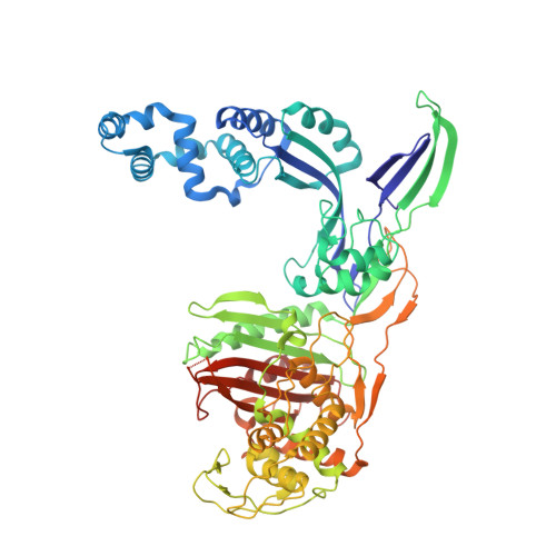
Crystal Structures of Penicillin-Binding Protein 3 (PBP3) from Methicillin-Resistant Staphylococcus aureus in the Apo and Cefotaxime-Bound Forms.
Yoshida, H., Kawai, F., Obayashi, E., Akashi, S., Roper, D.I., Tame, J.R., Park, S.Y.(2012) J Mol Biology 423: 351-364
- PubMed: 22846910
- DOI: https://doi.org/10.1016/j.jmb.2012.07.012
- Primary Citation of Related Structures:
3VSK, 3VSL - PubMed Abstract:
Staphylococcus aureus is a widespread Gram-positive opportunistic pathogen, and a methicillin-resistant form (MRSA) is particularly difficult to treat clinically. We have solved two crystal structures of penicillin-binding protein (PBP) 3 (PBP3) from MRSA, the apo form and a complex with the β-lactam antibiotic cefotaxime, and used electrospray mass spectrometry to measure its sensitivity to a variety of penicillin derivatives. PBP3 is a class B PBP, possessing an N-terminal non-penicillin-binding domain, sometimes called a dimerization domain, and a C-terminal transpeptidase domain. The model shows a different orientation of its two domains compared to earlier models of other class B PBPs and a novel, larger N-domain. Consistent with the nomenclature of "dimerization domain", the N-terminal region forms an apparently tight interaction with a neighboring molecule related by a 2-fold symmetry axis in the crystal structure. This dimer form is predicted to be highly stable in solution by the PISA server, but mass spectrometry and analytical ultracentrifugation provide unequivocal evidence that the protein is a monomer in solution.
- Protein Design Laboratory, Yokohama City University, Suehiro 1-7-29, Tsurumi-ku, Yokohama 230-0045, Japan.
Organizational Affiliation:


