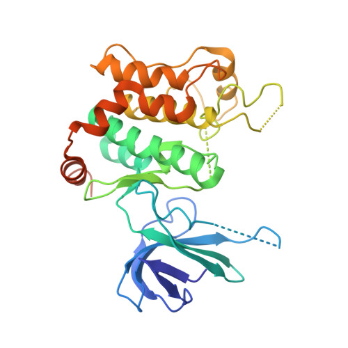
3WZU

THE STRUCTURE OF MAP2K7 IN COMPLEX WITH 5Z-7-oxozeaenol


Experimental Data Snapshot

  • Method: X-RAY DIFFRACTION
  • Resolution: 3.01 Å
  • R-Value Free: 0.322 
  • R-Value Work: 0.236 
  • R-Value Observed: 0.240 

Starting Model: experimental
View more details

wwPDB Validation   3D Report Full Report


Ligand Structure Quality Assessment 


This is version 1.3 of the entry. See complete history


Literature

5Z-7-Oxozeaenol covalently binds to MAP2K7 at Cys218 in an unprecedented manner.

Sogabe, Y.Matsumoto, T.Hashimoto, T.Kirii, Y.Sawa, M.Kinoshita, T.

(2015) Bioorg Med Chem Lett 25: 593-596

  • DOI: https://doi.org/10.1016/j.bmcl.2014.12.011
  • Primary Citation of Related Structures:  
    3WZU

  • PubMed Abstract: 

    5Z-7-Oxozeaenol (5Z7O) is a covalent bonding inhibitor against the several protein kinases (e.g., ERK2 and TAK1) that possess a free cysteine at the gatekeeper-2 position. In addition to this cysteine, MAP2K7 has three other cysteine residues that are candidate for covalent bonding by the inhibitor 5Z7O. The crystal structure of the MAP2K7/5Z7O complex revealed that the inhibitor binds to MAP2K7 at a cysteine residue located at the end of the hinge region and not at the gatekeeper-2 residue. The structural insights into the interaction of 5Z7O with MAP2K7 should aid the development of 5Z7O derivatives with improved potency and selectivity.


  • Organizational Affiliation

    Graduate School of Science, Osaka Prefecture University, 1-1 Gakuen-cho, Naka-ku, Sakai, Osaka 599-8531, Japan.


Macromolecules
Find similar proteins by:  (by identity cutoff)  |  3D Structure
Entity ID: 1
MoleculeChains Sequence LengthOrganismDetailsImage
Dual specificity mitogen-activated protein kinase kinase 7324Homo sapiensMutation(s): 0 
Gene Names: MAP2K7JNKK2MEK7MKK7PRKMK7SKK4
EC: 2.7.12.2
UniProt & NIH Common Fund Data Resources
Find proteins for O14733 (Homo sapiens)
Explore O14733 
Go to UniProtKB:  O14733
PHAROS:  O14733
GTEx:  ENSG00000076984 
Entity Groups  
Sequence Clusters30% Identity50% Identity70% Identity90% Identity95% Identity100% Identity
UniProt GroupO14733
Sequence Annotations
Expand
  • Reference Sequence
Small Molecules
Ligands 1 Unique
IDChains Name / Formula / InChI Key2D Diagram3D Interactions
1FM
Query on 1FM

Download Ideal Coordinates CCD File 
B [auth A](3S,5Z,8S,9S,11E)-8,9,16-trihydroxy-14-methoxy-3-methyl-3,4,9,10-tetrahydro-1H-2-benzoxacyclotetradecine-1,7(8H)-dione
C19 H22 O7
NEQZWEXWOFPKOT-BYRRXHGESA-N
Binding Affinity Annotations 
IDSourceBinding Affinity
1FM BindingDB:  3WZU IC50: 1300 (nM) from 1 assay(s)
Experimental Data & Validation

Experimental Data

  • Method: X-RAY DIFFRACTION
  • Resolution: 3.01 Å
  • R-Value Free: 0.322 
  • R-Value Work: 0.236 
  • R-Value Observed: 0.240 
  • Space Group: P 61 2 2
Unit Cell:
Length ( Å )Angle ( ˚ )
a = 71.395α = 90
b = 71.395β = 90
c = 268.817γ = 120
Software Package:
Software NamePurpose
ADSCdata collection
MOLREPphasing
REFMACrefinement
HKL-2000data reduction
SCALEPACKdata scaling

Structure Validation

View Full Validation Report


Ligand Structure Quality Assessment 


Entry History 

Deposition Data

Revision History  (Full details and data files)

  • Version 1.0: 2015-01-14
    Type: Initial release
  • Version 1.1: 2017-11-22
    Changes: Refinement description
  • Version 1.2: 2022-08-24
    Changes: Database references, Derived calculations
  • Version 1.3: 2023-11-08
    Changes: Data collection, Refinement description