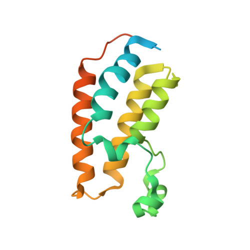
4A9I

N-TERMINAL BROMODOMAIN OF HUMAN BRD2 WITH 3-methyl-1,2,3,4- tetrahydroquinazolin-2-one


Experimental Data Snapshot

  • Method: X-RAY DIFFRACTION
  • Resolution: 2.25 Å
  • R-Value Free: 0.252 
  • R-Value Work: 0.195 
  • R-Value Observed: 0.197 

wwPDB Validation   3D Report Full Report


Ligand Structure Quality Assessment 


This is version 1.3 of the entry. See complete history


Literature

Fragment-Based Discovery of Bromodomain Inhibitors Part 1: Inhibitor Binding Modes and Implications for Lead Discovery.

Chung, C.W.Dean, A.W.Woolven, J.M.Bamborough, P.

(2012) J Med Chem 55: 576

  • DOI: https://doi.org/10.1021/jm201320w
  • Primary Citation of Related Structures:  
    4A9E, 4A9F, 4A9H, 4A9I, 4A9J, 4A9K, 4A9L

  • PubMed Abstract: 

    Bromodomain-containing proteins are key epigenetic regulators of gene transcription and readers of the histone code. However, the therapeutic benefits of modulating this target class are largely unexplored due to the lack of suitable chemical probes. This article describes the generation of lead molecules for the BET bromodomains through screening a fragment set chosen using structural insights and computational approaches. Analysis of 40 BRD2/fragment X-ray complexes highlights both shared and disparate interaction features that may be exploited for affinity and selectivity. Six representative crystal structures are then exemplified in detail. Two of the fragments are completely new bromodomain chemotypes, and three have never before been crystallized in a bromodomain, so our results significantly extend the limited public knowledge-base of crystallographic small molecule/bromodomain interactions. Certain fragments (including paracetamol) bind in a consistent mode to different bromodomains such as CREBBP, suggesting their potential to act as generic bromodomain templates. An important implication is that the bromodomains are not only a phylogenetic family but also a system in which chemical and structural knowledge of one bromodomain gives insights transferrable to others.


  • Organizational Affiliation

    Computational & Structural Chemistry, Molecular Discovery Research, GlaxoSmithKline R&D, Medicines Research Centre, Gunnels Wood Road, Stevenage, Hertfordshire SG1 2NY, UK. Chun-wa.chung@gsk.com


Macromolecules
Find similar proteins by:  (by identity cutoff)  |  3D Structure
Entity ID: 1
MoleculeChains Sequence LengthOrganismDetailsImage
BROMODOMAIN CONTAINING 2
A, B, C
154Homo sapiensMutation(s): 0 
UniProt & NIH Common Fund Data Resources
Find proteins for P25440 (Homo sapiens)
Explore P25440 
Go to UniProtKB:  P25440
PHAROS:  P25440
GTEx:  ENSG00000204256 
Entity Groups  
Sequence Clusters30% Identity50% Identity70% Identity90% Identity95% Identity100% Identity
UniProt GroupP25440
Sequence Annotations
Expand
  • Reference Sequence
Small Molecules
Binding Affinity Annotations 
IDSourceBinding Affinity
P9I PDBBind:  4A9I IC50: 1.26e+4 (nM) from 1 assay(s)
Experimental Data & Validation

Experimental Data

  • Method: X-RAY DIFFRACTION
  • Resolution: 2.25 Å
  • R-Value Free: 0.252 
  • R-Value Work: 0.195 
  • R-Value Observed: 0.197 
  • Space Group: C 1 2 1
Unit Cell:
Length ( Å )Angle ( ˚ )
a = 114.232α = 90
b = 55.531β = 94.85
c = 67.975γ = 90
Software Package:
Software NamePurpose
REFMACrefinement

Structure Validation

View Full Validation Report


Ligand Structure Quality Assessment 


Entry History 

Deposition Data

Revision History  (Full details and data files)

  • Version 1.0: 2012-01-11
    Type: Initial release
  • Version 1.1: 2012-02-08
    Changes: Other
  • Version 1.2: 2019-05-08
    Changes: Data collection, Experimental preparation, Other
  • Version 1.3: 2024-05-08
    Changes: Data collection, Database references, Derived calculations, Other