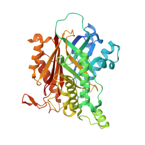
Cyanuric Acid Hydrolase: Evolutionary Innovation by Structural Concatenation.
Peat, T.S., Balotra, S., Wilding, M., French, N.G., Briggs, L.J., Panjikar, S., Cowieson, N., Newman, J., Scott, C.(2013) Mol Microbiol 88: 1149
- PubMed: 23651355
- DOI: https://doi.org/10.1111/mmi.12249
- Primary Citation of Related Structures:
4BVQ, 4BVR, 4BVS, 4BVT - PubMed Abstract:
The cyanuric acid hydrolase, AtzD, is the founding member of a newly identified family of ring-opening amidases. We report the first X-ray structure for this family, which is a novel fold (termed the 'Toblerone' fold) that likely evolved via the concatenation of monomers of the trimeric YjgF superfamily and the acquisition of a metal binding site. Structures of AtzD with bound substrate (cyanuric acid) and inhibitors (phosphate, barbituric acid and melamine), along with mutagenesis studies, allowed the identification of the active site. The AtzD monomer, active site and substrate all possess threefold rotational symmetry, to the extent that the active site possesses three potential Ser-Lys catalytic dyads. A single catalytic dyad (Ser85-Lys42) is hypothesized, based on biochemical evidence and crystallographic data. A plausible catalytic mechanism based on these observations is also presented. A comparison with a homology model of the related barbiturase, Bar, was used to infer the active-site residues responsible for substrate specificity, and the phylogeny of the 68 AtzD-like enzymes in the database were analysed in light of this structure-function relationship.
- CSIRO Materials, Science and Engineering, Parkville, Vic., 3801, Australia.
Organizational Affiliation:


