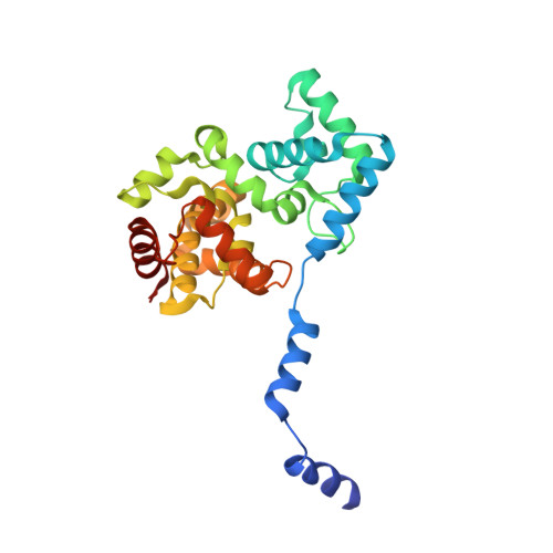
Structure of severe Fever with thrombocytopenia syndrome virus nucleocapsid protein in complex with suramin reveals therapeutic potential
Jiao, L., Ouyang, S., Liang, M., Niu, F., Shaw, N., Wu, W., Ding, W., Jin, C., Peng, Y., Zhu, Y., Zhang, F., Wang, T., Li, C., Zuo, X., Luan, C.H., Li, D., Liu, Z.J.(2013) J Virol 87: 6829-6839
- PubMed: 23576501
- DOI: https://doi.org/10.1128/JVI.00672-13
- Primary Citation of Related Structures:
4J4R, 4J4S, 4J4U, 4J4V, 4J4W, 4J4X, 4J4Y - PubMed Abstract:
Severe fever with thrombocytopenia syndrome is an emerging infectious disease caused by a novel bunyavirus (SFTSV). Lack of vaccines and inadequate therapeutic treatments have made the spread of the virus a global concern. Viral nucleocapsid protein (N) is essential for its transcription and replication. Here, we present the crystal structures of N from SFTSV and its homologs from Buenaventura (BUE) and Granada (GRA) viruses. The structures reveal that phleboviral N folds into a compact core domain and an extended N-terminal arm that mediates oligomerization, such as tetramer, pentamer, and hexamer of N assemblies. Structural superimposition indicates that phleboviral N adopts a conserved architecture and uses a similar RNA encapsidation strategy as that of RVFV-N. The RNA binding cavity runs along the inner edge of the ring-like assembly. A triple mutant of SFTSV-N, R64D/K67D/K74D, almost lost its ability to bind RNA in vitro, is deficient in its ability to transcribe and replicate. Structural studies of the mutant reveal that both alterations in quaternary assembly and the charge distribution contribute to the loss of RNA binding. In the screening of inhibitors Suramin was identified to bind phleboviral N specifically. The complex crystal structure of SFTSV-N with Suramin was refined to a 2.30-Å resolution. Suramin was found sitting in the putative RNA binding cavity of SFTSV-N. The inhibitory effect of Suramin on SFTSV replication was confirmed in Vero cells. Therefore, a common Suramin-based therapeutic approach targeting SFTSV-N and its homologs could be developed for containing phleboviral outbreaks.
Organizational Affiliation:
National Laboratory of Biomacromolecules, Institute of Biophysics, Chinese Academy of Sciences, Beijing, China.


