4J5H | pdb_00004j5h

Crystal Structure of B. thuringiensis AiiA mutant F107W with N-decanoyl-L-homoserine bound at the active site


Experimental Data Snapshot

  • Method: X-RAY DIFFRACTION
  • Resolution: 1.45 Å
  • R-Value Free: 
    0.151 (Depositor), 0.150 (DCC) 
  • R-Value Work: 
    0.120 (Depositor), 0.120 (DCC) 
  • R-Value Observed: 
    0.122 (Depositor) 

wwPDB Validation   3D Report Full Report


Ligand Structure Quality Assessment 

Created with Raphaël 2.3.0Worse 01 BetterLigand structure goodness of fit to experimental dataBest fitted 1K4Click on this verticalbar to view details

This is version 1.1 of the entry. See complete history


Literature

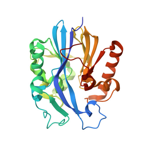
A phenylalanine clamp controls substrate specificity in the quorum-quenching metallo-gamma-lactonase from Bacillus thuringiensis.

Liu, C.F.Liu, D.Momb, J.Thomas, P.W.Lajoie, A.Petsko, G.A.Fast, W.Ringe, D.

(2013) Biochemistry 52: 1603-1610

  • DOI: https://doi.org/10.1021/bi400050j
  • Primary Citation of Related Structures:  
    4J5F, 4J5H

  • PubMed Abstract: 

    Autoinducer inactivator A (AiiA) is a metal-dependent N-acyl homoserine lactone hydrolase that displays broad substrate specificity but shows a preference for substrates with long N-acyl substitutions. Previously, crystal structures of AiiA in complex with the ring-opened product N-hexanoyl-l-homoserine revealed binding interactions near the metal center but did not identify a binding pocket for the N-acyl chains of longer substrates. Here we report the crystal structure of an AiiA mutant, F107W, determined in the presence and absence of N-decanoyl-l-homoserine. F107 is located in a hydrophobic cavity adjacent to the previously identified ligand binding pocket, and the F107W mutation results in the formation of an unexpected interaction with the ring-opened product. Notably, the structure reveals a previously unidentified hydrophobic binding pocket for the substrate's N-acyl chain. Two aromatic residues, F64 and F68, form a hydrophobic clamp, centered around the seventh carbon in the product-bound structure's decanoyl chain, making an interaction that would also be available for longer substrates, but not for shorter substrates. Steady-state kinetics using substrates of various lengths with AiiA bearing mutations at the hydrophobic clamp, including insertion of a redox-sensitive cysteine pair, confirms the importance of this hydrophobic feature for substrate preference. Identifying the specificity determinants of AiiA will aid the development of more selective quorum-quenching enzymes as tools and as potential therapeutics.


  • Organizational Affiliation

    Department of Chemistry and Rosenstiel Basic Medical Sciences Research Center, Brandeis University, Waltham, Massachusetts 02454, USA.


Macromolecules
Find similar proteins by:  (by identity cutoff)  |  3D Structure
Entity ID: 1
MoleculeChains Sequence LengthOrganismDetailsImage
N-acyl homoserine lactonase254Bacillus thuringiensisMutation(s): 1 
Gene Names: aiiA
EC: 3.1.1.81
UniProt
Find proteins for A3FJ64 (Bacillus thuringiensis)
Explore A3FJ64 
Go to UniProtKB:  A3FJ64
Entity Groups  
Sequence Clusters30% Identity50% Identity70% Identity90% Identity95% Identity100% Identity
UniProt GroupA3FJ64
Sequence Annotations
Expand
  • Reference Sequence
Small Molecules
Ligands 3 Unique
IDChains Name / Formula / InChI Key2D Diagram3D Interactions
1K4
Query on 1K4

Download Ideal Coordinates CCD File 
D [auth A]N-decanoyl-L-homoserine
C14 H27 N O4
SRVJGOTYLHNRPN-LBPRGKRZSA-N
GOL
Query on GOL

Download Ideal Coordinates CCD File 
E [auth A]
F [auth A]
G [auth A]
H [auth A]
I [auth A]
GLYCEROL
C3 H8 O3
PEDCQBHIVMGVHV-UHFFFAOYSA-N
ZN
Query on ZN

Download Ideal Coordinates CCD File 
B [auth A],
C [auth A]
ZINC ION
Zn
PTFCDOFLOPIGGS-UHFFFAOYSA-N
Experimental Data & Validation

Experimental Data

  • Method: X-RAY DIFFRACTION
  • Resolution: 1.45 Å
  • R-Value Free:  0.151 (Depositor), 0.150 (DCC) 
  • R-Value Work:  0.120 (Depositor), 0.120 (DCC) 
  • R-Value Observed: 0.122 (Depositor) 
Space Group: P 21 21 21
Unit Cell:
Length ( Å )Angle ( ˚ )
a = 54.825α = 90
b = 55.444β = 90
c = 80.396γ = 90
Software Package:
Software NamePurpose
DENZOdata reduction
SCALEPACKdata scaling
PHASERphasing
REFMACrefinement
PDB_EXTRACTdata extraction

Structure Validation

View Full Validation Report


Ligand Structure Quality Assessment 

Created with Raphaël 2.3.0Worse 01 BetterLigand structure goodness of fit to experimental dataBest fitted 1K4Click on this verticalbar to view details

Entry History 

Deposition Data

Revision History  (Full details and data files)

  • Version 1.0: 2013-06-26
    Type: Initial release
  • Version 1.1: 2024-02-28
    Changes: Data collection, Database references, Derived calculations