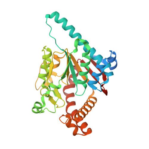
4NE2

Pantothenamide-bound Pantothenate Kinase from Klebsiella pneumoniae


Experimental Data Snapshot

  • Method: X-RAY DIFFRACTION
  • Resolution: 1.90 Å
  • R-Value Free: 0.214 
  • R-Value Work: 0.154 
  • R-Value Observed: 0.157 

Starting Model: experimental
View more details

wwPDB Validation   3D Report Full Report


Ligand Structure Quality Assessment 


This is version 1.2 of the entry. See complete history


Literature

Structural characterization of a new N-substituted pantothenamide bound to pantothenate kinases from Klebsiella pneumoniae and Staphylococcus aureus.

Hughes, S.J.Antoshchenko, T.Kim, K.P.Smil, D.Park, H.W.

(2014) Proteins 82: 1542-1548

  • DOI: https://doi.org/10.1002/prot.24524
  • Primary Citation of Related Structures:  
    4NB4, 4NE2

  • PubMed Abstract: 

    Pantothenate kinase (PanK) is the rate-limiting enzyme in Coenzyme A biosynthesis, catalyzing the ATP-dependent phosphorylation of pantothenate. We solved the co-crystal structures of PanKs from Staphylococcus aureus (SaPanK) and Klebsiella pneumonia (KpPanK) with N-[2-(1,3-benzodioxol-5-yl)ethyl] pantothenamide (N354-Pan). Two different N354-Pan conformers interact with polar/nonpolar mixed residues in SaPanK and aromatic residues in KpPanK. Additionally, phosphorylated N354-Pan is found at the closed active site of SaPanK but not at the open active site of KpPanK, suggesting an exchange of the phosphorylated product with a new N354-Pan only in KpPanK. Together, pantothenamides conformational flexibility and binding pocket are two key considerations for selective compound design.


  • Organizational Affiliation

    Department of Pharmacology, University of Toronto, Toronto, Ontario, M5G 1L7.


Macromolecules
Find similar proteins by:  (by identity cutoff)  |  3D Structure
Entity ID: 1
MoleculeChains Sequence LengthOrganismDetailsImage
Pantothenate kinase
A, B
334Klebsiella pneumoniae 342Mutation(s): 0 
Gene Names: coaAKPK_5321UUU_44690
EC: 2.7.1.33
UniProt
Find proteins for B5XYG3 (Klebsiella pneumoniae (strain 342))
Explore B5XYG3 
Go to UniProtKB:  B5XYG3
Entity Groups  
Sequence Clusters30% Identity50% Identity70% Identity90% Identity95% Identity100% Identity
UniProt GroupB5XYG3
Sequence Annotations
Expand
  • Reference Sequence
Small Molecules
Ligands 3 Unique
IDChains Name / Formula / InChI Key2D Diagram3D Interactions
ADP
Query on ADP

Download Ideal Coordinates CCD File 
C [auth A],
F [auth B]
ADENOSINE-5'-DIPHOSPHATE
C10 H15 N5 O10 P2
XTWYTFMLZFPYCI-KQYNXXCUSA-N
SH2
Query on SH2

Download Ideal Coordinates CCD File 
D [auth A],
G [auth B]
(R)-N-(3-((2-(benzo[d][1,3]dioxol-5-yl)ethyl)amino)-3-oxopropyl)-2,4-dihydroxy-3,3-dimethylbutanamide
C18 H26 N2 O6
WTOXXQQUHIAOIP-INIZCTEOSA-N
MG
Query on MG

Download Ideal Coordinates CCD File 
E [auth A],
H [auth B]
MAGNESIUM ION
Mg
JLVVSXFLKOJNIY-UHFFFAOYSA-N
Experimental Data & Validation

Experimental Data

  • Method: X-RAY DIFFRACTION
  • Resolution: 1.90 Å
  • R-Value Free: 0.214 
  • R-Value Work: 0.154 
  • R-Value Observed: 0.157 
  • Space Group: F 2 2 2
Unit Cell:
Length ( Å )Angle ( ˚ )
a = 106.765α = 90
b = 180.366β = 90
c = 181.488γ = 90
Software Package:
Software NamePurpose
SCALEPACKdata scaling
PHASERphasing
REFMACrefinement
PDB_EXTRACTdata extraction
DENZOdata reduction

Structure Validation

View Full Validation Report


Ligand Structure Quality Assessment 


Entry History 

Deposition Data

Revision History  (Full details and data files)

  • Version 1.0: 2014-02-26
    Type: Initial release
  • Version 1.1: 2014-06-25
    Changes: Database references
  • Version 1.2: 2023-09-20
    Changes: Data collection, Database references, Derived calculations, Refinement description