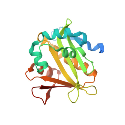
4NXY

Crystal Structure of the GNAT domain of S. lividans PAT


Experimental Data Snapshot

  • Method: X-RAY DIFFRACTION
  • Resolution: 1.45 Å
  • R-Value Free: 0.217 
  • R-Value Work: 0.176 
  • R-Value Observed: 0.178 

wwPDB Validation   3D Report Full Report


This is version 1.3 of the entry. See complete history


Literature

Insights into the specificity of lysine acetyltransferases.

Tucker, A.C.Taylor, K.C.Rank, K.C.Rayment, I.Escalante-Semerena, J.C.

(2014) J Biol Chem 289: 36249-36262

  • DOI: https://doi.org/10.1074/jbc.M114.613901
  • Primary Citation of Related Structures:  
    4NXY, 4U5Y

  • PubMed Abstract: 

    Reversible lysine acetylation by protein acetyltransferases is a conserved regulatory mechanism that controls diverse cellular pathways. Gcn5-related N-acetyltransferases (GNATs), named after their founding member, are found in all domains of life. GNATs are known for their role as histone acetyltransferases, but non-histone bacterial protein acetytransferases have been identified. Only structures of GNAT complexes with short histone peptide substrates are available in databases. Given the biological importance of this modification and the abundance of lysine in polypeptides, how specificity is attained for larger protein substrates is central to understanding acetyl-lysine-regulated networks. Here we report the structure of a GNAT in complex with a globular protein substrate solved to 1.9 Å. GNAT binds the protein substrate with extensive surface interactions distinct from those reported for GNAT-peptide complexes. Our data reveal determinants needed for the recognition of a protein substrate and provide insight into the specificity of GNATs.


  • Organizational Affiliation

    From the Department of Microbiology, University of Georgia, Athens, Georgia 30602 and.


Macromolecules
Find similar proteins by:  (by identity cutoff)  |  3D Structure
Entity ID: 1
MoleculeChains Sequence LengthOrganismDetailsImage
Acyl-CoA synthetase194Streptomyces lividans TK24Mutation(s): 0 
Gene Names: SSPG_01886
Entity Groups  
Sequence Clusters30% Identity50% Identity70% Identity90% Identity95% Identity100% Identity
Sequence Annotations
Expand
  • Reference Sequence
Small Molecules
Ligands 2 Unique
IDChains Name / Formula / InChI Key2D Diagram3D Interactions
ETF
Query on ETF

Download Ideal Coordinates CCD File 
B [auth A]TRIFLUOROETHANOL
C2 H3 F3 O
RHQDFWAXVIIEBN-UHFFFAOYSA-N
GOL
Query on GOL

Download Ideal Coordinates CCD File 
C [auth A]GLYCEROL
C3 H8 O3
PEDCQBHIVMGVHV-UHFFFAOYSA-N
Experimental Data & Validation

Experimental Data

  • Method: X-RAY DIFFRACTION
  • Resolution: 1.45 Å
  • R-Value Free: 0.217 
  • R-Value Work: 0.176 
  • R-Value Observed: 0.178 
  • Space Group: P 21 21 21
Unit Cell:
Length ( Å )Angle ( ˚ )
a = 40.056α = 90
b = 56.711β = 90
c = 74γ = 90
Software Package:
Software NamePurpose
HKL-3000data collection
HKL-3000phasing
REFMACrefinement
HKL-3000data reduction
HKL-3000data scaling

Structure Validation

View Full Validation Report


Entry History 

Deposition Data

Revision History  (Full details and data files)

  • Version 1.0: 2014-11-19
    Type: Initial release
  • Version 1.1: 2014-11-26
    Changes: Database references
  • Version 1.2: 2015-01-14
    Changes: Database references
  • Version 1.3: 2024-02-28
    Changes: Data collection, Database references, Derived calculations