4P9H

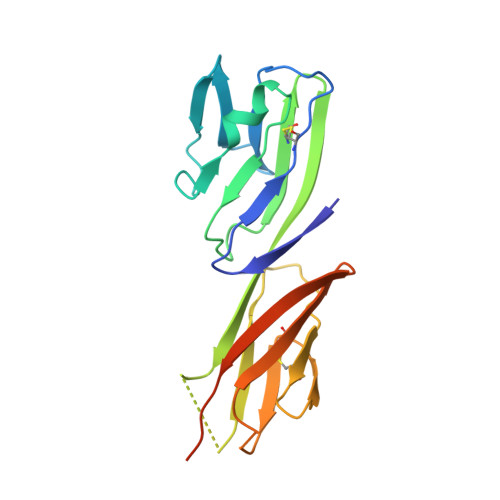
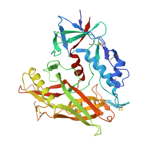
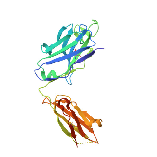
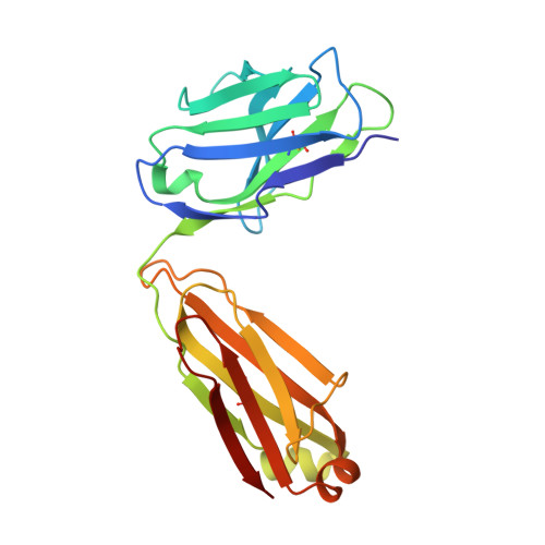
Crystal structure of 8ANC195 Fab in complex with gp120 of 93TH057 HIV-1 and soluble CD4 D1D2


Experimental Data Snapshot

  • Method: X-RAY DIFFRACTION
  • Resolution: 3.00 Å
  • R-Value Free: 0.272 
  • R-Value Work: 0.235 
  • R-Value Observed: 0.237 

Starting Models: experimental
View more details

wwPDB Validation   3D Report Full Report


Ligand Structure Quality Assessment 


This is version 4.2 of the entry. See complete history


Literature

Antibody 8ANC195 Reveals a Site of Broad Vulnerability on the HIV-1 Envelope Spike.

Scharf, L.Scheid, J.F.Lee, J.H.West, A.P.Chen, C.Gao, H.Gnanapragasam, P.N.Mares, R.Seaman, M.S.Ward, A.B.Nussenzweig, M.C.Bjorkman, P.J.

(2014) Cell Rep 7: 785-795

  • DOI: https://doi.org/10.1016/j.celrep.2014.04.001
  • Primary Citation of Related Structures:  
    4P9H, 4P9M

  • PubMed Abstract: 

    Broadly neutralizing antibodies (bNAbs) to HIV-1 envelope glycoprotein (Env) can prevent infection in animal models. Characterized bNAb targets, although key to vaccine and therapeutic strategies, are currently limited. We defined a new site of vulnerability by solving structures of bNAb 8ANC195 complexed with monomeric gp120 by X-ray crystallography and trimeric Env by electron microscopy. The site includes portions of gp41 and N-linked glycans adjacent to the CD4-binding site on gp120, making 8ANC195 the first donor-derived anti-HIV-1 bNAb with an epitope spanning both Env subunits. Rather than penetrating the glycan shield by using a single variable-region CDR loop, 8ANC195 inserted its entire heavy-chain variable domain into a gap to form a large interface with gp120 glycans and regions of the gp120 inner domain not contacted by other bNAbs. By isolating additional 8ANC195 clonal variants, we identified a more potent variant, which may be valuable for therapeutic approaches using bNAb combinations with nonoverlapping epitopes.


  • Organizational Affiliation

    Division of Biology and Biological Engineering, California Institute of Technology, Pasadena, CA 91125, USA.


Macromolecules
Find similar proteins by:  (by identity cutoff)  |  3D Structure
Entity ID: 1
MoleculeChains Sequence LengthOrganismDetailsImage
T-cell surface glycoprotein CD4A [auth C]192Homo sapiensMutation(s): 0 
Gene Names: CD4
UniProt & NIH Common Fund Data Resources
Find proteins for P01730 (Homo sapiens)
Explore P01730 
Go to UniProtKB:  P01730
PHAROS:  P01730
GTEx:  ENSG00000010610 
Entity Groups  
Sequence Clusters30% Identity50% Identity70% Identity90% Identity95% Identity100% Identity
UniProt GroupP01730
Glycosylation
Glycosylation Sites: 1Go to GlyGen: P01730-1
Sequence Annotations
Expand
  • Reference Sequence
Find similar proteins by:  (by identity cutoff)  |  3D Structure
Entity ID: 2
MoleculeChains Sequence LengthOrganismDetailsImage
Envelope glycoprotein gp160B [auth G]361Human immunodeficiency virus 1Mutation(s): 0 
Gene Names: env
UniProt
Find proteins for Q0ED31 (Human immunodeficiency virus 1)
Explore Q0ED31 
Go to UniProtKB:  Q0ED31
Entity Groups  
Sequence Clusters30% Identity50% Identity70% Identity90% Identity95% Identity100% Identity
UniProt GroupQ0ED31
Glycosylation
Glycosylation Sites: 7
Sequence Annotations
Expand
  • Reference Sequence
Find similar proteins by:  (by identity cutoff)  |  3D Structure
Entity ID: 3
MoleculeChains Sequence LengthOrganismDetailsImage
Fab heavy chainC [auth H]244Homo sapiensMutation(s): 0 
Entity Groups  
Sequence Clusters30% Identity50% Identity70% Identity90% Identity95% Identity100% Identity
Glycosylation
Glycosylation Sites: 1
Sequence Annotations
Expand
  • Reference Sequence
Find similar proteins by:  (by identity cutoff)  |  3D Structure
Entity ID: 4
MoleculeChains Sequence LengthOrganismDetailsImage
Fab light chainD [auth L]215Homo sapiensMutation(s): 0 
Entity Groups  
Sequence Clusters30% Identity50% Identity70% Identity90% Identity95% Identity100% Identity
Sequence Annotations
Expand
  • Reference Sequence
Oligosaccharides

Help

Entity ID: 5
MoleculeChains Length2D Diagram Glycosylation3D Interactions
2-acetamido-2-deoxy-beta-D-glucopyranose-(1-4)-2-acetamido-2-deoxy-beta-D-glucopyranoseE [auth A],
I [auth F]
2N-Glycosylation
Glycosylation Resources
GlyTouCan:  G42666HT
GlyCosmos:  G42666HT
GlyGen:  G42666HT
Entity ID: 6
MoleculeChains Length2D Diagram Glycosylation3D Interactions
alpha-D-mannopyranose-(1-2)-alpha-D-mannopyranose-(1-2)-alpha-D-mannopyranose-(1-3)-[alpha-D-mannopyranose-(1-2)-alpha-D-mannopyranose-(1-6)-[alpha-D-mannopyranose-(1-3)]alpha-D-mannopyranose-(1-6)]beta-D-mannopyranose-(1-4)-2-acetamido-2-deoxy-beta-D-glucopyranose-(1-4)-2-acetamido-2-deoxy-beta-D-glucopyranoseF [auth B]10N-Glycosylation
Glycosylation Resources
GlyTouCan:  G40702WU
GlyCosmos:  G40702WU
GlyGen:  G40702WU
Entity ID: 7
MoleculeChains Length2D Diagram Glycosylation3D Interactions
alpha-D-mannopyranose-(1-3)-beta-D-mannopyranose-(1-4)-2-acetamido-2-deoxy-beta-D-glucopyranose-(1-4)-2-acetamido-2-deoxy-beta-D-glucopyranoseG [auth D]4N-Glycosylation
Glycosylation Resources
GlyTouCan:  G81315DD
GlyCosmos:  G81315DD
GlyGen:  G81315DD
Entity ID: 8
MoleculeChains Length2D Diagram Glycosylation3D Interactions
alpha-D-mannopyranose-(1-3)-[alpha-D-mannopyranose-(1-6)]beta-D-mannopyranose-(1-4)-2-acetamido-2-deoxy-beta-D-glucopyranose-(1-4)-2-acetamido-2-deoxy-beta-D-glucopyranoseH [auth E]5N-Glycosylation
Glycosylation Resources
GlyTouCan:  G22768VO
GlyCosmos:  G22768VO
GlyGen:  G22768VO
Entity ID: 9
MoleculeChains Length2D Diagram Glycosylation3D Interactions
alpha-D-mannopyranose-(1-6)-beta-D-mannopyranose-(1-4)-2-acetamido-2-deoxy-beta-D-glucopyranose-(1-4)-2-acetamido-2-deoxy-beta-D-glucopyranoseJ [auth I]4N-Glycosylation
Glycosylation Resources
GlyTouCan:  G22573RC
GlyCosmos:  G22573RC
GlyGen:  G22573RC
Small Molecules
Ligands 3 Unique
IDChains Name / Formula / InChI Key2D Diagram3D Interactions
EPE
Query on EPE

Download Ideal Coordinates CCD File 
K [auth G]4-(2-HYDROXYETHYL)-1-PIPERAZINE ETHANESULFONIC ACID
C8 H18 N2 O4 S
JKMHFZQWWAIEOD-UHFFFAOYSA-N
NAG
Query on NAG

Download Ideal Coordinates CCD File 
N [auth G],
O [auth G],
P [auth H]
2-acetamido-2-deoxy-beta-D-glucopyranose
C8 H15 N O6
OVRNDRQMDRJTHS-FMDGEEDCSA-N
BEN
Query on BEN

Download Ideal Coordinates CCD File 
L [auth G],
M [auth G]
BENZAMIDINE
C7 H8 N2
PXXJHWLDUBFPOL-UHFFFAOYSA-N
Experimental Data & Validation

Experimental Data

  • Method: X-RAY DIFFRACTION
  • Resolution: 3.00 Å
  • R-Value Free: 0.272 
  • R-Value Work: 0.235 
  • R-Value Observed: 0.237 
  • Space Group: P 21 21 21
Unit Cell:
Length ( Å )Angle ( ˚ )
a = 66.529α = 90
b = 132.492β = 90
c = 142.769γ = 90
Software Package:
Software NamePurpose
PHENIXrefinement
XDSdata reduction
PDB_EXTRACTdata extraction
PHASERphasing
Aimlessdata scaling

Structure Validation

View Full Validation Report


Ligand Structure Quality Assessment 


Entry History 

Deposition Data

Revision History  (Full details and data files)

  • Version 1.0: 2014-05-21
    Type: Initial release
  • Version 2.0: 2017-11-22
    Changes: Atomic model, Derived calculations, Refinement description, Source and taxonomy, Structure summary
  • Version 3.0: 2020-07-29
    Type: Remediation
    Reason: Carbohydrate remediation
    Changes: Atomic model, Data collection, Derived calculations, Structure summary
  • Version 3.1: 2021-03-24
    Changes: Source and taxonomy, Structure summary
  • Version 4.0: 2021-08-04
    Changes: Atomic model, Data collection, Derived calculations, Non-polymer description, Structure summary
  • Version 4.1: 2023-09-27
    Changes: Data collection, Database references, Refinement description
  • Version 4.2: 2024-10-23
    Changes: Structure summary