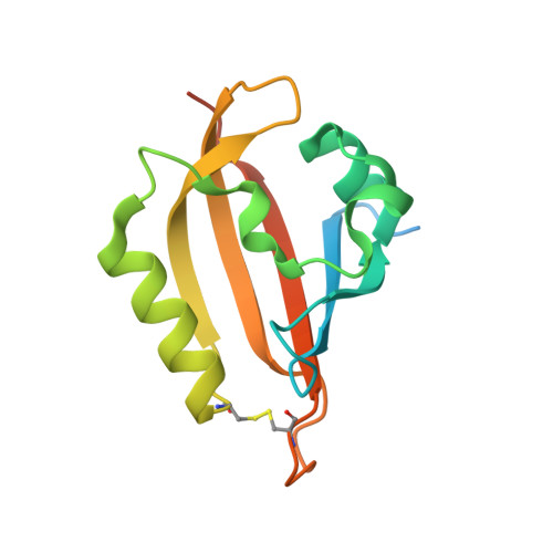
Full-length structure of a monomeric histidine kinase reveals basis for sensory regulation.
Rivera-Cancel, G., Ko, W.H., Tomchick, D.R., Correa, F., Gardner, K.H.(2014) Proc Natl Acad Sci U S A 111: 17839-17844
- PubMed: 25468971
- DOI: https://doi.org/10.1073/pnas.1413983111
- Primary Citation of Related Structures:
4R38, 4R39, 4R3A - PubMed Abstract:
Although histidine kinases (HKs) are critical sensors of external stimuli in prokaryotes, the mechanisms by which their sensor domains control enzymatic activity remain unclear. Here, we report the full-length structure of a blue light-activated HK from Erythrobacter litoralis HTCC2594 (EL346) and the results of biochemical and biophysical studies that explain how it is activated by light. Contrary to the standard view that signaling occurs within HK dimers, EL346 functions as a monomer. Its structure reveals that the light-oxygen-voltage (LOV) sensor domain both controls kinase activity and prevents dimerization by binding one side of a dimerization/histidine phosphotransfer-like (DHpL) domain. The DHpL domain also contacts the catalytic/ATP-binding (CA) domain, keeping EL346 in an inhibited conformation in the dark. Upon light stimulation, interdomain interactions weaken to facilitate activation. Our data suggest that the LOV domain controls kinase activity by affecting the stability of the DHpL/CA interface, releasing the CA domain from an inhibited conformation upon photoactivation. We suggest parallels between EL346 and dimeric HKs, with sensor-induced movements in the DHp similarly remodeling the DHp/CA interface as part of activation.
- Departments of Biophysics and Biochemistry, University of Texas Southwestern Medical Center, Dallas, TX 75390-8816;
Organizational Affiliation:


