4WX4

Crystal structure of adenovirus 8 protease in complex with a nitrile inhibitor


Experimental Data Snapshot

  • Method: X-RAY DIFFRACTION
  • Resolution: 1.03 Å
  • R-Value Free: 0.137 
  • R-Value Work: 0.117 
  • R-Value Observed: 0.118 

wwPDB Validation   3D Report Full Report


Ligand Structure Quality Assessment 


This is version 1.1 of the entry. See complete history


Literature

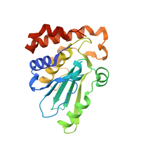
Structure-based design and optimization of potent inhibitors of the adenoviral protease.

Grosche, P.Sirockin, F.Mac Sweeney, A.Ramage, P.Erbel, P.Melkko, S.Bernardi, A.Hughes, N.Ellis, D.Combrink, K.D.Jarousse, N.Altmann, E.

(2015) Bioorg Med Chem Lett 25: 438-443

  • DOI: https://doi.org/10.1016/j.bmcl.2014.12.057
  • Primary Citation of Related Structures:  
    4WX4, 4WX6, 4WX7

  • PubMed Abstract: 

    Adenoviral infections are associated with a wide range of acute diseases, among which ocular viral conjunctivitis (EKC) and disseminated disease in immunocompromised patients. To date, no approved specific anti-adenoviral drug is available, but there is a growing need for an effective treatment of such infections. The adenoviral protease, adenain, plays a crucial role for the viral lifecycle and thus represents an attractive therapeutic target. Structure-guided design with the objective to depeptidize tetrapeptide nitrile 1 led to the novel chemotype 2. Optimization of scaffold 2 resulted in picomolar adenain inhibitors 3a and 3b. In addition, a complementary series of irreversible vinyl sulfone containing inhibitors were rationally designed, prepared and evaluated against adenoviral protease. High resolution X-ray co-crystal structures of representatives of each series proves the successful design of these inhibitors and provides an excellent basis for future medicinal chemistry optimization of these compounds.


  • Organizational Affiliation

    Novartis Institute for Biomedical Research, Novartis Campus, CH-4002 Basel, Switzerland.


Macromolecules
Find similar proteins by:  (by identity cutoff)  |  3D Structure
Entity ID: 1
MoleculeChains Sequence LengthOrganismDetailsImage
Protease205Human adenovirus D8Mutation(s): 0 
UniProt
Find proteins for B9A5C1 (Human adenovirus D serotype 8)
Explore B9A5C1 
Go to UniProtKB:  B9A5C1
Entity Groups  
Sequence Clusters30% Identity50% Identity70% Identity90% Identity95% Identity100% Identity
UniProt GroupB9A5C1
Sequence Annotations
Expand
  • Reference Sequence

Find similar proteins by:  Sequence   |   3D Structure  

Entity ID: 2
MoleculeChains Sequence LengthOrganismDetailsImage
peptideB [auth C]10synthetic constructMutation(s): 0 
Entity Groups  
Sequence Clusters30% Identity50% Identity70% Identity90% Identity95% Identity100% Identity
Sequence Annotations
Expand
  • Reference Sequence
Small Molecules
Ligands 3 Unique
IDChains Name / Formula / InChI Key2D Diagram3D Interactions
3VF
Query on 3VF

Download Ideal Coordinates CCD File 
C [auth A]N-[(2-cyanopyrimidin-4-yl)methyl]-3-[2-(3,5-dichlorophenyl)-2-methylpropanoyl]-4-methoxybenzamide
C24 H20 Cl2 N4 O3
WCQQOAFZCDVMEP-UHFFFAOYSA-N
EPE
Query on EPE

Download Ideal Coordinates CCD File 
D [auth A]4-(2-HYDROXYETHYL)-1-PIPERAZINE ETHANESULFONIC ACID
C8 H18 N2 O4 S
JKMHFZQWWAIEOD-UHFFFAOYSA-N
GLY
Query on GLY

Download Ideal Coordinates CCD File 
E [auth C]GLYCINE
C2 H5 N O2
DHMQDGOQFOQNFH-UHFFFAOYSA-N
Experimental Data & Validation

Experimental Data

  • Method: X-RAY DIFFRACTION
  • Resolution: 1.03 Å
  • R-Value Free: 0.137 
  • R-Value Work: 0.117 
  • R-Value Observed: 0.118 
  • Space Group: P 1 21 1
Unit Cell:
Length ( Å )Angle ( ˚ )
a = 41.454α = 90
b = 42.79β = 93.16
c = 59.059γ = 90
Software Package:
Software NamePurpose
XSCALEdata scaling
REFMACrefinement
PDB_EXTRACTdata extraction
XDSdata reduction
PHASERphasing

Structure Validation

View Full Validation Report


Ligand Structure Quality Assessment 


Entry History 

Revision History  (Full details and data files)

  • Version 1.0: 2015-01-14
    Type: Initial release
  • Version 1.1: 2015-01-28
    Changes: Database references