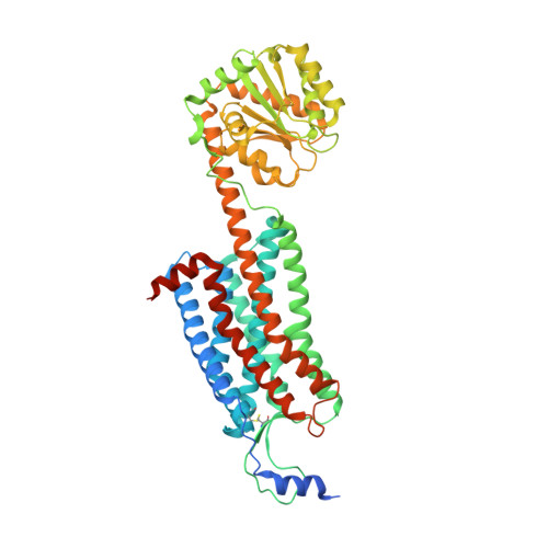
4ZJ8

Structures of the human OX1 orexin receptor bound to selective and dual antagonists


Experimental Data Snapshot

  • Method: X-RAY DIFFRACTION
  • Resolution: 2.75 Å
  • R-Value Free: 0.273 
  • R-Value Work: 0.223 
  • R-Value Observed: 0.225 

Starting Models: experimental
View more details

wwPDB Validation   3D Report Full Report


Ligand Structure Quality Assessment 


This is version 1.4 of the entry. See complete history


Literature

Structure and ligand-binding mechanism of the human OX1 and OX2 orexin receptors.

Yin, J.Babaoglu, K.Brautigam, C.A.Clark, L.Shao, Z.Scheuermann, T.H.Harrell, C.M.Gotter, A.L.Roecker, A.J.Winrow, C.J.Renger, J.J.Coleman, P.J.Rosenbaum, D.M.

(2016) Nat Struct Mol Biol 23: 293-299

  • DOI: https://doi.org/10.1038/nsmb.3183
  • Primary Citation of Related Structures:  
    4ZJ8, 4ZJC

  • PubMed Abstract: 

    The orexin (also known as hypocretin) G protein-coupled receptors (GPCRs) regulate sleep and other behavioral functions in mammals, and are therapeutic targets for sleep and wake disorders. The human receptors hOX1R and hOX2R, which are 64% identical in sequence, have overlapping but distinct physiological functions and potential therapeutic profiles. We determined structures of hOX1R bound to the OX1R-selective antagonist SB-674042 and the dual antagonist suvorexant at 2.8-Å and 2.75-Å resolution, respectively, and used molecular modeling to illuminate mechanisms of antagonist subtype selectivity between hOX1R and hOX2R. The hOX1R structures also reveal a conserved amphipathic α-helix, in the extracellular N-terminal region, that interacts with orexin-A and is essential for high-potency neuropeptide activation at both receptors. The orexin-receptor crystal structures are valuable tools for the design and development of selective orexin-receptor antagonists and agonists.


  • Organizational Affiliation

    Department of Biophysics, University of Texas Southwestern Medical Center, Dallas, Texas, USA.


Macromolecules
Find similar proteins by:  (by identity cutoff)  |  3D Structure
Entity ID: 1
MoleculeChains Sequence LengthOrganismDetailsImage
human OX1R fusion protein to P.abysii glycogen synthase553Homo sapiensPyrococcus abyssi GE5Mutation(s): 1 
Gene Names: HCRTR1PAB2292PYRAB00770
Membrane Entity: Yes 
UniProt & NIH Common Fund Data Resources
Find proteins for Q9V2J8 (Pyrococcus abyssi (strain GE5 / Orsay))
Explore Q9V2J8 
Go to UniProtKB:  Q9V2J8
Find proteins for O43613 (Homo sapiens)
Explore O43613 
Go to UniProtKB:  O43613
PHAROS:  O43613
GTEx:  ENSG00000121764 
Entity Groups  
Sequence Clusters30% Identity50% Identity70% Identity90% Identity95% Identity100% Identity
UniProt GroupsQ9V2J8O43613
Sequence Annotations
Expand
  • Reference Sequence
Small Molecules
Ligands 2 Unique
IDChains Name / Formula / InChI Key2D Diagram3D Interactions
SUV
Query on SUV

Download Ideal Coordinates CCD File 
B [auth A][(7R)-4-(5-chloro-1,3-benzoxazol-2-yl)-7-methyl-1,4-diazepan-1-yl][5-methyl-2-(2H-1,2,3-triazol-2-yl)phenyl]methanone
C23 H23 Cl N6 O2
JYTNQNCOQXFQPK-MRXNPFEDSA-N
OLA
Query on OLA

Download Ideal Coordinates CCD File 
C [auth A]
D [auth A]
E [auth A]
F [auth A]
G [auth A]
C [auth A],
D [auth A],
E [auth A],
F [auth A],
G [auth A],
H [auth A]
OLEIC ACID
C18 H34 O2
ZQPPMHVWECSIRJ-KTKRTIGZSA-N
Binding Affinity Annotations 
IDSourceBinding Affinity
SUV BindingDB:  4ZJ8 Ki: min: 0.4, max: 8.7 (nM) from 6 assay(s)
IC50: min: 50, max: 199 (nM) from 2 assay(s)
Experimental Data & Validation

Experimental Data

  • Method: X-RAY DIFFRACTION
  • Resolution: 2.75 Å
  • R-Value Free: 0.273 
  • R-Value Work: 0.223 
  • R-Value Observed: 0.225 
  • Space Group: P 21 21 21
Unit Cell:
Length ( Å )Angle ( ˚ )
a = 63.242α = 90
b = 64.992β = 90
c = 181.478γ = 90
Software Package:
Software NamePurpose
PHENIXrefinement
Blu-Icedata collection
HKL-3000data scaling
PHASERphasing
HKL-3000data reduction

Structure Validation

View Full Validation Report


Ligand Structure Quality Assessment 


Entry History 

Deposition Data

Revision History  (Full details and data files)

  • Version 1.0: 2016-03-09
    Type: Initial release
  • Version 1.1: 2016-03-23
    Changes: Database references
  • Version 1.2: 2016-04-13
    Changes: Database references
  • Version 1.3: 2023-09-27
    Changes: Data collection, Database references, Derived calculations, Refinement description
  • Version 1.4: 2024-10-23
    Changes: Structure summary