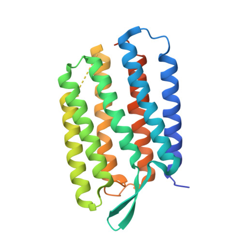
5A44

Structure of Bacteriorhodopsin obtained from 20um crystals by multi crystal data collection


Experimental Data Snapshot

  • Method: X-RAY DIFFRACTION
  • Resolution: 2.29 Å
  • R-Value Free: 0.239 
  • R-Value Work: 0.232 
  • R-Value Observed: 0.232 

wwPDB Validation   3D Report Full Report


Ligand Structure Quality Assessment 


This is version 1.1 of the entry. See complete history


Literature

Meshandcollect: An Automated Multi-Crystal Data-Collection Workflow for Synchrotron Macromolecular Crystallography Beamlines.

Zander, U.Bourenkov, G.Popov, A.N.De Sanctis, D.Svensson, O.Mccarthy, A.A.Round, E.Gordeliy, V.Mueller-Dieckmann, C.Leonard, G.A.

(2015) Acta Crystallogr D Biol Crystallogr 71: 2328

  • DOI: https://doi.org/10.1107/S1399004715017927
  • Primary Citation of Related Structures:  
    5A3Y, 5A3Z, 5A44, 5A45, 5A47

  • PubMed Abstract: 

    Here, an automated procedure is described to identify the positions of many cryocooled crystals mounted on the same sample holder, to rapidly predict and rank their relative diffraction strengths and to collect partial X-ray diffraction data sets from as many of the crystals as desired. Subsequent hierarchical cluster analysis then allows the best combination of partial data sets, optimizing the quality of the final data set obtained. The results of applying the method developed to various systems and scenarios including the compilation of a complete data set from tiny crystals of the membrane protein bacteriorhodopsin and the collection of data sets for successful structure determination using the single-wavelength anomalous dispersion technique are also presented.


  • Organizational Affiliation

    Structural Biology Group, European Synchrotron Radiation Facility, CS 40220, 38043 Grenoble, France.


Macromolecules
Find similar proteins by:  (by identity cutoff)  |  3D Structure
Entity ID: 1
MoleculeChains Sequence LengthOrganismDetailsImage
BACTERIORHODOPSIN248Halobacterium salinarumMutation(s): 0 
Membrane Entity: Yes 
UniProt
Find proteins for P02945 (Halobacterium salinarum (strain ATCC 700922 / JCM 11081 / NRC-1))
Explore P02945 
Go to UniProtKB:  P02945
Entity Groups  
Sequence Clusters30% Identity50% Identity70% Identity90% Identity95% Identity100% Identity
UniProt GroupP02945
Sequence Annotations
Expand
  • Reference Sequence
Small Molecules
Ligands 3 Unique
IDChains Name / Formula / InChI Key2D Diagram3D Interactions
LI1
Query on LI1

Download Ideal Coordinates CCD File 
C [auth A]
D [auth A]
E [auth A]
F [auth A]
G [auth A]
C [auth A],
D [auth A],
E [auth A],
F [auth A],
G [auth A],
H [auth A],
I [auth A],
J [auth A],
K [auth A]
1-[2,6,10.14-TETRAMETHYL-HEXADECAN-16-YL]-2-[2,10,14-TRIMETHYLHEXADECAN-16-YL]GLYCEROL
C42 H86 O3
YERVUJAKCNBGCR-BIHSMRAKSA-N
RET
Query on RET

Download Ideal Coordinates CCD File 
B [auth A]RETINAL
C20 H28 O
NCYCYZXNIZJOKI-OVSJKPMPSA-N
HP6
Query on HP6

Download Ideal Coordinates CCD File 
L [auth A]HEPTANE
C7 H16
IMNFDUFMRHMDMM-UHFFFAOYSA-N
Experimental Data & Validation

Experimental Data

  • Method: X-RAY DIFFRACTION
  • Resolution: 2.29 Å
  • R-Value Free: 0.239 
  • R-Value Work: 0.232 
  • R-Value Observed: 0.232 
  • Space Group: P 63
Unit Cell:
Length ( Å )Angle ( ˚ )
a = 61.127α = 90
b = 61.127β = 90
c = 110.313γ = 120
Software Package:
Software NamePurpose
REFMACrefinement

Structure Validation

View Full Validation Report


Ligand Structure Quality Assessment 


Entry History 

Revision History  (Full details and data files)

  • Version 1.0: 2015-11-11
    Type: Initial release
  • Version 1.1: 2024-11-13
    Changes: Data collection, Database references, Derived calculations, Other, Structure summary