5C80

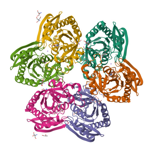
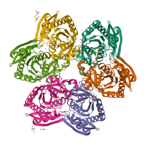
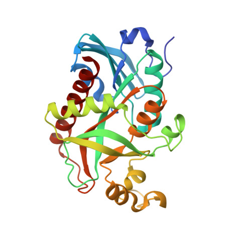
X-ray structure uridine phosphorylase from Vibrio cholerae in complex with uridine at 2.24 A resolution


Experimental Data Snapshot

  • Method: X-RAY DIFFRACTION
  • Resolution: 2.24 Å
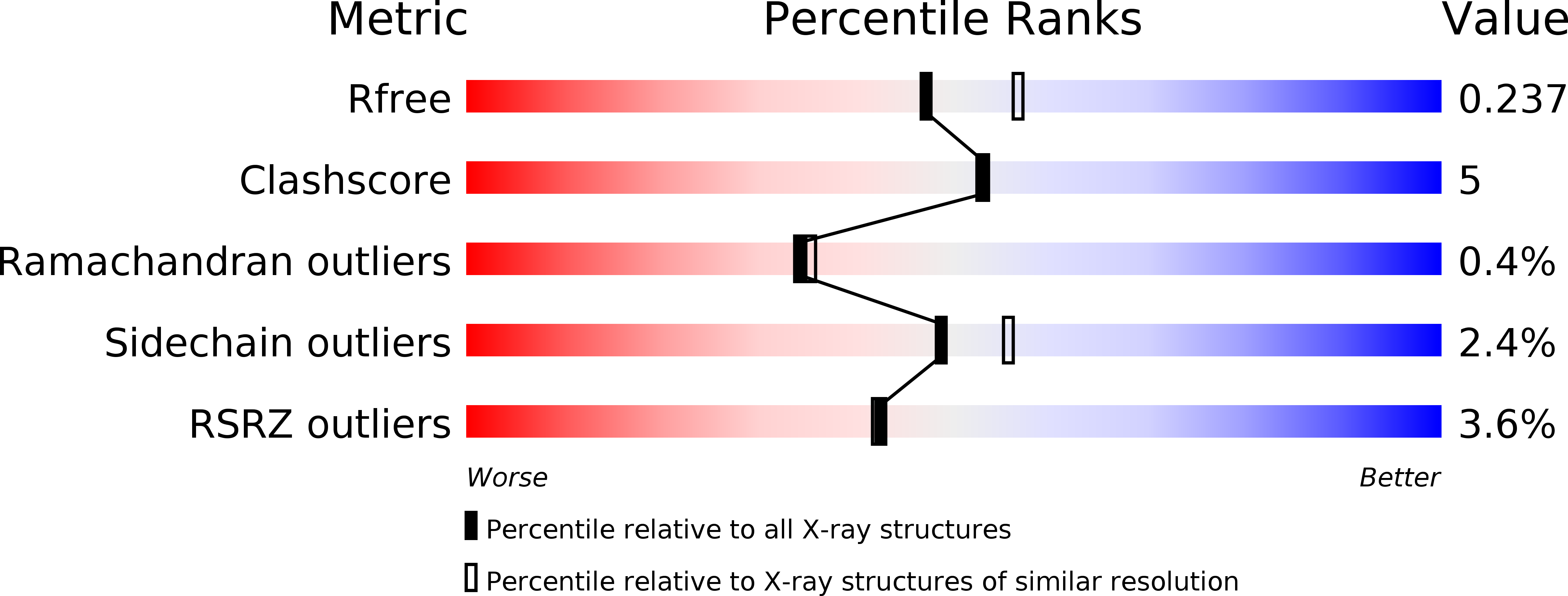
  • R-Value Free: 0.237 
  • R-Value Work: 0.168 
  • R-Value Observed: 0.172 

Starting Model: experimental
View more details

wwPDB Validation   3D Report Full Report


Ligand Structure Quality Assessment 

Created with Raphaël 2.3.0Worse 01 BetterLigand structure goodness of fit to experimental dataBest fitted URIClick on this verticalbar to view details

This is version 1.2 of the entry. See complete history


Literature

X-ray structures of uridine phosphorylase from Vibrio cholerae in complexes with uridine, thymidine, uracil, thymine, and phosphate anion: Substrate specificity of bacterial uridine phosphorylases

Prokofev, I.I.Lashkov, A.A.Gabdoulkhakov, A.G.Balaev, V.V.Seregina, T.A.Mironov, A.S.Betzel, C.Mikhailov, A.M.

(2016) Crystallogr Rep 61: 954-973


Macromolecules
Find similar proteins by:  (by identity cutoff)  |  3D Structure
Entity ID: 1
MoleculeChains Sequence LengthOrganismDetailsImage
Uridine phosphorylase
A, B, C, D, E
253Vibrio choleraeMutation(s): 0 
Gene Names: udpDN30_1909VC39_02535VC78_02550VS27_10630WG08_05660
EC: 2.4.2.3
UniProt
Find proteins for Q9K4U1 (Vibrio cholerae)
Explore Q9K4U1 
Go to UniProtKB:  Q9K4U1
Entity Groups  
Sequence Clusters30% Identity50% Identity70% Identity90% Identity95% Identity100% Identity
UniProt GroupQ9K4U1
Sequence Annotations
Expand
  • Reference Sequence
Small Molecules
Ligands 6 Unique
IDChains Name / Formula / InChI Key2D Diagram3D Interactions
URI
Query on URI

Download Ideal Coordinates CCD File 
AA [auth E]
CA [auth F]
G [auth A]
L [auth B]
R [auth C]
URIDINE
C9 H12 N2 O6
DRTQHJPVMGBUCF-XVFCMESISA-N
TRS
Query on TRS

Download Ideal Coordinates CCD File 
BA [auth E]
DA [auth F]
EA [auth F]
FA [auth F]
H [auth A]
2-AMINO-2-HYDROXYMETHYL-PROPANE-1,3-DIOL
C4 H12 N O3
LENZDBCJOHFCAS-UHFFFAOYSA-O
PEG
Query on PEG

Download Ideal Coordinates CCD File 
I [auth A],
O [auth B],
T [auth C]
DI(HYDROXYETHYL)ETHER
C4 H10 O3
MTHSVFCYNBDYFN-UHFFFAOYSA-N
GOL
Query on GOL

Download Ideal Coordinates CCD File 
GA [auth F],
N [auth B],
Y [auth D]
GLYCEROL
C3 H8 O3
PEDCQBHIVMGVHV-UHFFFAOYSA-N
CL
Query on CL

Download Ideal Coordinates CCD File 
HA [auth F]
J [auth A]
K [auth A]
P [auth B]
U [auth C]
CHLORIDE ION
Cl
VEXZGXHMUGYJMC-UHFFFAOYSA-M
NA
Query on NA

Download Ideal Coordinates CCD File 
IA [auth F],
Q [auth B]
SODIUM ION
Na
FKNQFGJONOIPTF-UHFFFAOYSA-N
Experimental Data & Validation

Experimental Data

  • Method: X-RAY DIFFRACTION
  • Resolution: 2.24 Å
  • R-Value Free: 0.237 
  • R-Value Work: 0.168 
  • R-Value Observed: 0.172 
  • Space Group: P 1
Unit Cell:
Length ( Å )Angle ( ˚ )
a = 61.255α = 71.81
b = 73.11β = 77.19
c = 83.137γ = 85.69
Software Package:
Software NamePurpose
SCALAdata scaling
PHASERphasing
REFMACrefinement
PDB_EXTRACTdata extraction

Structure Validation

View Full Validation Report


Ligand Structure Quality Assessment 

Created with Raphaël 2.3.0Worse 01 BetterLigand structure goodness of fit to experimental dataBest fitted URIClick on this verticalbar to view details

Entry History & Funding Information

Deposition Data


Funding OrganizationLocationGrant Number
RFBRRussian Federation14-04-00952a

Revision History  (Full details and data files)

  • Version 1.0: 2016-07-20
    Type: Initial release
  • Version 1.1: 2016-12-28
    Changes: Database references
  • Version 1.2: 2024-01-10
    Changes: Data collection, Database references, Refinement description