Complexity of the Ruminococcus Flavefaciens Cellulosome Reflects an Expansion in Glycan Recognition.
Venditto, I., Luis, A.S., Rydahl, M., Schuckel, J., Fernandes, V.O., Vidal-Melgosa, S., Bule, P., Goyal, A., Pires, V.M.R., Dourado, C.G., Ferreira, L.M.A., Coutinho, P.M., Henrissat, B., Knox, J.P., Basle, A., Najmudin, S., Gilbert, H.J., Willats, W.G.T., Fontes, C.M.G.A.(2016) Proc Natl Acad Sci U S A 113: 7136
- PubMed: 27298375
- DOI: https://doi.org/10.1073/pnas.1601558113
- Primary Citation of Related Structures:
4D3L, 4V17, 4V18, 4V1B, 4V1I, 4V1K, 4V1L, 5AOS, 5AOT, 5FU2, 5FU3, 5FU4, 5FU5 - PubMed Abstract:
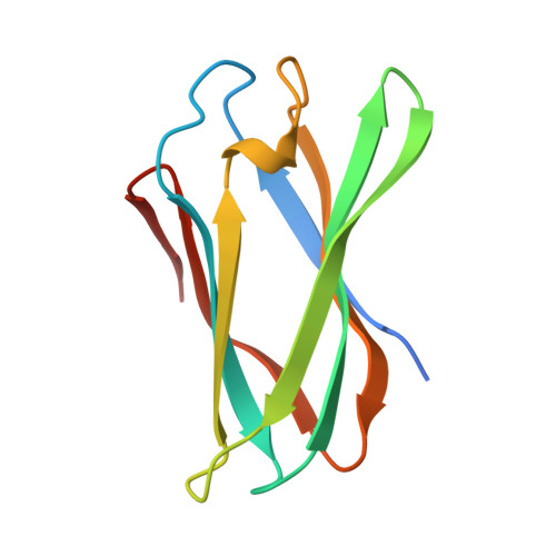
The breakdown of plant cell wall (PCW) glycans is an important biological and industrial process. Noncatalytic carbohydrate binding modules (CBMs) fulfill a critical targeting function in PCW depolymerization. Defining the portfolio of CBMs, the CBMome, of a PCW degrading system is central to understanding the mechanisms by which microbes depolymerize their target substrates. Ruminococcus flavefaciens, a major PCW degrading bacterium, assembles its catalytic apparatus into a large multienzyme complex, the cellulosome. Significantly, bioinformatic analyses of the R. flavefaciens cellulosome failed to identify a CBM predicted to bind to crystalline cellulose, a key feature of the CBMome of other PCW degrading systems. Here, high throughput screening of 177 protein modules of unknown function was used to determine the complete CBMome of R. flavefaciens The data identified six previously unidentified CBM families that targeted β-glucans, β-mannans, and the pectic polysaccharide homogalacturonan. The crystal structures of four CBMs, in conjunction with site-directed mutagenesis, provide insight into the mechanism of ligand recognition. In the CBMs that recognize β-glucans and β-mannans, differences in the conformation of conserved aromatic residues had a significant impact on the topology of the ligand binding cleft and thus ligand specificity. A cluster of basic residues in CBM77 confers calcium-independent recognition of homogalacturonan, indicating that the carboxylates of galacturonic acid are key specificity determinants. This report shows that the extended repertoire of proteins in the cellulosome of R. flavefaciens contributes to an extended CBMome that supports efficient PCW degradation in the absence of CBMs that specifically target crystalline cellulose.
Organizational Affiliation:
Interdisciplinary Centre of Research in Animal Health, Faculdade de Medicina Veterinária, Universidade de Lisboa, Pólo Universitário do Alto da Ajuda, 1300-477 Lisbon, Portugal; Institute for Cell and Molecular Biosciences, Newcastle University, Newcastle upon Tyne NE2 4HH, United Kingdom;


