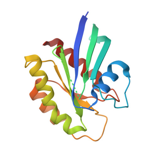
5VBZ

Crystal Structure of Small Molecule Disulfide 2C07 Bound to H-Ras M72C GppNHp


Experimental Data Snapshot

  • Method: X-RAY DIFFRACTION
  • Resolution: 2.20 Å
  • R-Value Free: 0.253 
  • R-Value Work: 0.210 
  • R-Value Observed: 0.212 

Starting Model: experimental
View more details

wwPDB Validation   3D Report Full Report


Ligand Structure Quality Assessment 


This is version 1.3 of the entry. See complete history


Literature

Ras Binder Induces a Modified Switch-II Pocket in GTP and GDP States.

Gentile, D.R.Rathinaswamy, M.K.Jenkins, M.L.Moss, S.M.Siempelkamp, B.D.Renslo, A.R.Burke, J.E.Shokat, K.M.

(2017) Cell Chem Biol 24: 1455-1466.e14

  • DOI: https://doi.org/10.1016/j.chembiol.2017.08.025
  • Primary Citation of Related Structures:  
    5VBE, 5VBM, 5VBZ

  • PubMed Abstract: 

    Covalent inhibitors of K-Ras(G12C) have been reported that exclusively recognize the GDP state. Here, we utilize disulfide tethering of a non-natural cysteine (K-Ras(M72C)) to identify a new switch-II pocket (S-IIP) binding ligand (2C07) that engages the active GTP state. Co-crystal structures of 2C07 bound to H-Ras(M72C) reveal binding in a cryptic groove we term S-IIG. In the GppNHp state, 2C07 binding to a modified S-IIP pushes switch I away from the nucleotide, breaking the network of polar contacts essential for adopting the canonical GTP state. Biochemical studies show that 2C07 alters nucleotide preference and inhibits SOS binding and catalyzed nucleotide exchange. 2C07 was converted to irreversible covalent analogs, which target both nucleotide states, inhibit PI3K activation in vitro, and function as occupancy probes to detect reversible engagement in competition assays. Targeting both nucleotide states opens the possibility of inhibiting oncogenic mutants of Ras, which exist predominantly in the GTP state in cells.


  • Organizational Affiliation

    Department of Cellular and Molecular Pharmacology, Howard Hughes Medical Institute, University of California, San Francisco, CA 94158, USA.


Macromolecules
Find similar proteins by:  (by identity cutoff)  |  3D Structure
Entity ID: 1
MoleculeChains Sequence LengthOrganismDetailsImage
GTPase HRas
A, B, C
171Homo sapiensMutation(s): 1 
Gene Names: HRASHRAS1
EC: 3.6.5.2
UniProt & NIH Common Fund Data Resources
Find proteins for P01112 (Homo sapiens)
Explore P01112 
Go to UniProtKB:  P01112
PHAROS:  P01112
GTEx:  ENSG00000174775 
Entity Groups  
Sequence Clusters30% Identity50% Identity70% Identity90% Identity95% Identity100% Identity
UniProt GroupP01112
Sequence Annotations
Expand
  • Reference Sequence
Experimental Data & Validation

Experimental Data

  • Method: X-RAY DIFFRACTION
  • Resolution: 2.20 Å
  • R-Value Free: 0.253 
  • R-Value Work: 0.210 
  • R-Value Observed: 0.212 
  • Space Group: P 21 21 21
Unit Cell:
Length ( Å )Angle ( ˚ )
a = 61.84α = 90
b = 75.44β = 90
c = 93.63γ = 90
Software Package:
Software NamePurpose
SCALAdata scaling
PHENIXrefinement
PDB_EXTRACTdata extraction
iMOSFLMdata reduction
PHASERphasing

Structure Validation

View Full Validation Report


Ligand Structure Quality Assessment 


Entry History 

Deposition Data

Revision History  (Full details and data files)

  • Version 1.0: 2017-10-25
    Type: Initial release
  • Version 1.1: 2017-11-01
    Changes: Database references
  • Version 1.2: 2018-01-03
    Changes: Database references
  • Version 1.3: 2023-10-04
    Changes: Data collection, Database references, Derived calculations, Refinement description