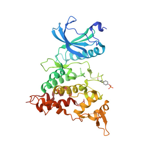
6A1F

Crystal structure of human DYRK1A in complex with compound 14


Experimental Data Snapshot

  • Method: X-RAY DIFFRACTION
  • Resolution: 1.50 Å
  • R-Value Free: 0.195 
  • R-Value Work: 0.177 
  • R-Value Observed: 0.178 

Starting Model: experimental
View more details

wwPDB Validation   3D Report Full Report


Ligand Structure Quality Assessment 


This is version 1.3 of the entry. See complete history


Literature

Discovery of DS42450411 as a potent orally active hepcidin production inhibitor: Design and optimization of novel 4-aminopyrimidine derivatives.

Fukuda, T.Ishiyama, T.Katagiri, T.Ueda, K.Muramatsu, S.Hashimoto, M.Aki, A.Baba, D.Watanabe, K.Tanaka, N.

(2018) Bioorg Med Chem Lett 28: 3333-3337

  • DOI: https://doi.org/10.1016/j.bmcl.2018.09.010
  • Primary Citation of Related Structures:  
    6A1F, 6A1G

  • PubMed Abstract: 

    Hepcidin has emerged as the central regulatory molecule in systemic iron homeostasis. The inhibition of hepcidin may be a favorable strategy for the treatment of anemia of chronic disease. Here, we have reported the design, synthesis, and structure-activity relationships (SAR) of a series of 4-aminopyrimidine compounds as inhibitors of hepcidin production. The optimization study of 1 led to the design of a potent and bioavailable inhibitor of hepcidin production, 34 (DS42450411), which showed serum hepcidin-lowering effects in a mouse model of interleukin-6-induced acute inflammation.


  • Organizational Affiliation

    Rare Disease Laboratories, Daiichi Sankyo Co., Ltd, 1-2-58 Hiromachi, Shinagawa-ku, Tokyo 140-8710, Japan. Electronic address: fukuda.takeshi.zv@daiichisankyo.co.jp.


Macromolecules
Find similar proteins by:  (by identity cutoff)  |  3D Structure
Entity ID: 1
MoleculeChains Sequence LengthOrganismDetailsImage
Dual specificity tyrosine-phosphorylation-regulated kinase 1A360Homo sapiensMutation(s): 0 
Gene Names: DYRK1ADYRKMNBMNBH
EC: 2.7.12.1 (PDB Primary Data), 2.7.11.23 (UniProt)
UniProt & NIH Common Fund Data Resources
Find proteins for Q13627 (Homo sapiens)
Explore Q13627 
Go to UniProtKB:  Q13627
PHAROS:  Q13627
GTEx:  ENSG00000157540 
Entity Groups  
Sequence Clusters30% Identity50% Identity70% Identity90% Identity95% Identity100% Identity
UniProt GroupQ13627
Sequence Annotations
Expand
  • Reference Sequence
Small Molecules
Ligands 3 Unique
IDChains Name / Formula / InChI Key2D Diagram3D Interactions
9OF (Subject of Investigation/LOI)
Query on 9OF

Download Ideal Coordinates CCD File 
H [auth A]8-methoxy-5,5-dimethyl-5,6-dihydrobenzo[h]quinazolin-4-amine
C15 H17 N3 O
JWHJIHHTDSIYQK-UHFFFAOYSA-N
PGE
Query on PGE

Download Ideal Coordinates CCD File 
I [auth A]TRIETHYLENE GLYCOL
C6 H14 O4
ZIBGPFATKBEMQZ-UHFFFAOYSA-N
SO4
Query on SO4

Download Ideal Coordinates CCD File 
B [auth A]
C [auth A]
D [auth A]
E [auth A]
F [auth A]
B [auth A],
C [auth A],
D [auth A],
E [auth A],
F [auth A],
G [auth A]
SULFATE ION
O4 S
QAOWNCQODCNURD-UHFFFAOYSA-L
Modified Residues  1 Unique
IDChains TypeFormula2D DiagramParent
PTR
Query on PTR
A
L-PEPTIDE LINKINGC9 H12 N O6 PTYR
Binding Affinity Annotations 
IDSourceBinding Affinity
9OF BindingDB:  6A1F IC50: 330 (nM) from 1 assay(s)
Experimental Data & Validation

Experimental Data

  • Method: X-RAY DIFFRACTION
  • Resolution: 1.50 Å
  • R-Value Free: 0.195 
  • R-Value Work: 0.177 
  • R-Value Observed: 0.178 
  • Space Group: C 1 2 1
Unit Cell:
Length ( Å )Angle ( ˚ )
a = 99.341α = 90
b = 69.806β = 117.65
c = 67.332γ = 90
Software Package:
Software NamePurpose
REFMACrefinement
HKL-2000data reduction
SCALEPACKdata scaling
PHASERphasing

Structure Validation

View Full Validation Report


Ligand Structure Quality Assessment 


Entry History 

Deposition Data

Revision History  (Full details and data files)

  • Version 1.0: 2018-10-03
    Type: Initial release
  • Version 1.1: 2018-10-24
    Changes: Data collection, Database references
  • Version 1.2: 2023-11-22
    Changes: Data collection, Database references, Refinement description
  • Version 1.3: 2024-10-16
    Changes: Structure summary