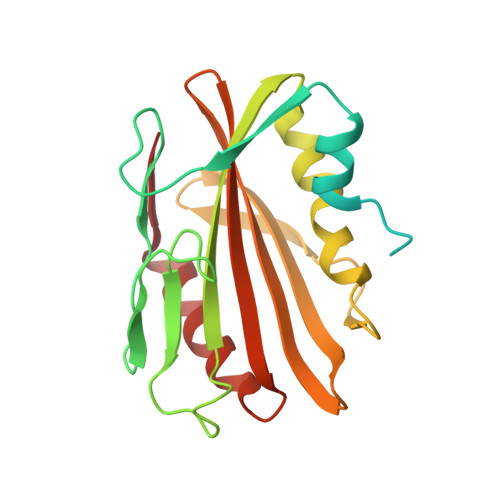
Structural and functional evidence that lipoprotein LpqN supports cell envelope biogenesis inMycobacterium tuberculosis.
Melly, G.C., Stokas, H., Dunaj, J.L., Hsu, F.F., Rajavel, M., Su, C.C., Yu, E.W., Purdy, G.E.(2019) J Biol Chem 294: 15711-15723
- PubMed: 31471317
- DOI: https://doi.org/10.1074/jbc.RA119.008781
- Primary Citation of Related Structures:
6E5D, 6E5F, 6MNA - PubMed Abstract:
The mycobacterial cell envelope is crucial to host-pathogen interactions as a barrier against antibiotics and the host immune response. In addition, cell envelope lipids are mycobacterial virulence factors. Cell envelope lipid biosynthesis is the target of a number of frontline tuberculosis treatments and has been the focus of much research. However, the transport mechanisms by which these lipids reach the mycomembrane remain poorly understood. Many envelope lipids are exported from the cytoplasm to the periplasmic space via the mycobacterial membrane protein large (MmpL) family of proteins. In other bacteria, lipoproteins can contribute to outer membrane biogenesis through direct binding of substrates and/or protein-protein associations with extracytoplasmic biosynthetic enzymes. In this report, we investigate whether the lipoprotein LpqN plays a similar role in mycobacteria. Using a genetic two-hybrid approach, we demonstrate that LpqN interacts with periplasmic loop domains of the MmpL3 and MmpL11 transporters that export mycolic acid-containing cell envelope lipids. We observe that LpqN also interacts with secreted cell envelope biosynthetic enzymes such as Ag85A via pulldown assays. The X-ray crystal structures of LpqN and LpqN bound to dodecyl-trehalose suggest that LpqN directly binds trehalose monomycolate, the MmpL3 and Ag85A substrate. Finally, we observe altered lipid profiles of the Δ lpqN mutant during biofilm maturation, pointing toward a possible physiological role for the protein. The results of this study suggest that LpqN may act as a membrane fusion protein, connecting MmpL transporters with periplasmic proteins, and provide general insight into the role of lipoproteins in Mycobacterium tuberculosis cell envelope biogenesis.
Organizational Affiliation:
Department of Molecular Microbiology & Immunology, Oregon Health & Science University, Portland, Oregon 97239.


