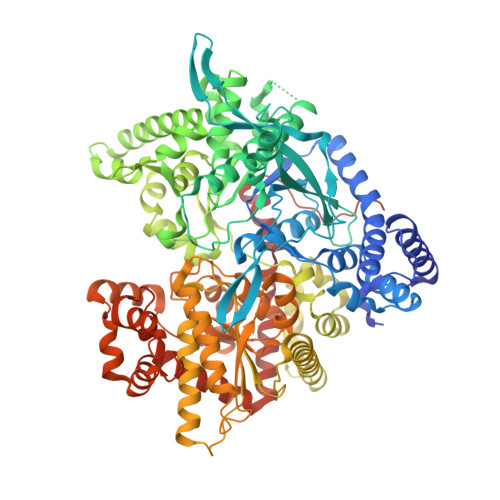
A multidisciplinary study of 3-( beta-d-glucopyranosyl)-5-substituted-1,2,4-triazole derivatives as glycogen phosphorylase inhibitors: Computation, synthesis, crystallography and kinetics reveal new potent inhibitors.
Kun, S., Begum, J., Kyriakis, E., Stamati, E.C.V., Barkas, T.A., Szennyes, E., Bokor, E., Szabo, K.E., Stravodimos, G.A., Sipos, A., Docsa, T., Gergely, P., Moffatt, C., Patraskaki, M.S., Kokolaki, M.C., Gkerdi, A., Skamnaki, V.T., Leonidas, D.D., Somsak, L., Hayes, J.M.(2018) Eur J Med Chem 147: 266-278
- PubMed: 29453094
- DOI: https://doi.org/10.1016/j.ejmech.2018.01.095
- Primary Citation of Related Structures:
6F3J, 6F3L, 6F3R, 6F3S, 6F3U - PubMed Abstract:
3-(β-d-Glucopyranosyl)-5-substituted-1,2,4-triazoles have been revealed as an effective scaffold for the development of potent glycogen phosphorylase (GP) inhibitors but with the potency very sensitive to the nature of the alkyl/aryl 5-substituent (Kun et al., Eur. J. Med. Chem. 2014, 76, 567). For a training set of these ligands, quantum mechanics-polarized ligand docking (QM-PLD) demonstrated good potential to identify larger differences in potencies (predictive index PI = 0.82) and potent inhibitors with K i 's < 10 μM (AU-ROC = 0.86). Accordingly, in silico screening of 2335 new analogues exploiting the ZINC docking database was performed and nine predicted candidates selected for synthesis. The compounds were prepared in O-perbenzoylated forms by either ring transformation of 5-β-d-glucopyranosyl tetrazole by N-benzyl-arenecarboximidoyl chlorides, ring closure of C-(β-d-glucopyranosyl)formamidrazone with aroyl chlorides, or that of N-(β-d-glucopyranosylcarbonyl)arenethiocarboxamides by hydrazine, followed by deprotections. Kinetics experiments against rabbit muscle GPb (rmGPb) and human liver GPa (hlGPa) revealed five compounds as potent low μM inhibitors with three of these on the submicromolar range for rmGPa. X-ray crystallographic analysis sourced the potency to a combination of favorable interactions from the 1,2,4-triazole and suitable aryl substituents in the GP catalytic site. The compounds also revealed promising calculated pharmacokinetic profiles.
Organizational Affiliation:
Department of Organic Chemistry, University of Debrecen, POB 400, H-4002 Debrecen, Hungary.


