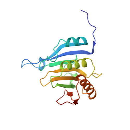
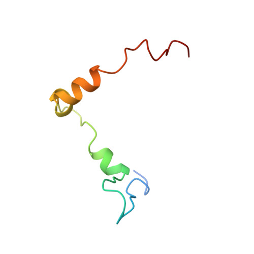
6FC2

Crystal structure of the eIF4E-Eap1p complex from Saccharomyces cerevisiae


Experimental Data Snapshot

  • Method: X-RAY DIFFRACTION
  • Resolution: 1.92 Å
  • R-Value Free: 0.206 
  • R-Value Work: 0.173 
  • R-Value Observed: 0.175 

Starting Model: experimental
View more details

wwPDB Validation   3D Report Full Report


This is version 1.2 of the entry. See complete history


Literature

Structural motifs in eIF4G and 4E-BPs modulate their binding to eIF4E to regulate translation initiation in yeast.

Gruner, S.Weber, R.Peter, D.Chung, M.Y.Igreja, C.Valkov, E.Izaurralde, E.

(2018) Nucleic Acids Res 46: 6893-6908

  • DOI: https://doi.org/10.1093/nar/gky542
  • Primary Citation of Related Structures:  
    6FBZ, 6FC0, 6FC1, 6FC2, 6FC3

  • PubMed Abstract: 

    The interaction of the eukaryotic initiation factor 4G (eIF4G) with the cap-binding protein eIF4E initiates cap-dependent translation and is regulated by the 4E-binding proteins (4E-BPs), which compete with eIF4G to repress translation. Metazoan eIF4G and 4E-BPs interact with eIF4E via canonical and non-canonical motifs that bind to the dorsal and lateral surface of eIF4E in a bipartite recognition mode. However, previous studies pointed to mechanistic differences in how fungi and metazoans regulate protein synthesis. We present crystal structures of the yeast eIF4E bound to two yeast 4E-BPs, p20 and Eap1p, as well as crystal structures of a fungal eIF4E-eIF4G complex. We demonstrate that the core principles of molecular recognition of eIF4E are in fact highly conserved among translational activators and repressors in eukaryotes. Finally, we reveal that highly specialized structural motifs do exist and serve to modulate the affinity of protein-protein interactions that regulate cap-dependent translation initiation in fungi.


  • Organizational Affiliation

    Department of Biochemistry, Max Planck Institute for Developmental Biology, Max-Planck-Ring 5, D-72076 Tübingen, Germany.


Macromolecules
Find similar proteins by:  (by identity cutoff)  |  3D Structure
Entity ID: 1
MoleculeChains Sequence LengthOrganismDetailsImage
Eukaryotic translation initiation factor 4E
A, C
183Saccharomyces cerevisiae S288CMutation(s): 3 
Gene Names: CDC33TIF45YOL139C
UniProt
Find proteins for P07260 (Saccharomyces cerevisiae (strain ATCC 204508 / S288c))
Explore P07260 
Go to UniProtKB:  P07260
Entity Groups  
Sequence Clusters30% Identity50% Identity70% Identity90% Identity95% Identity100% Identity
UniProt GroupP07260
Sequence Annotations
Expand
  • Reference Sequence
Find similar proteins by:  (by identity cutoff)  |  3D Structure
Entity ID: 2
MoleculeChains Sequence LengthOrganismDetailsImage
Protein EAP1
B, D
64Saccharomyces cerevisiae S288CMutation(s): 0 
Gene Names: EAP1YKL204W
UniProt
Find proteins for P36041 (Saccharomyces cerevisiae (strain ATCC 204508 / S288c))
Explore P36041 
Go to UniProtKB:  P36041
Entity Groups  
Sequence Clusters30% Identity50% Identity70% Identity90% Identity95% Identity100% Identity
UniProt GroupP36041
Sequence Annotations
Expand
  • Reference Sequence
Small Molecules
Ligands 1 Unique
IDChains Name / Formula / InChI Key2D Diagram3D Interactions
CA
Query on CA

Download Ideal Coordinates CCD File 
E [auth A]CALCIUM ION
Ca
BHPQYMZQTOCNFJ-UHFFFAOYSA-N
Experimental Data & Validation

Experimental Data

  • Method: X-RAY DIFFRACTION
  • Resolution: 1.92 Å
  • R-Value Free: 0.206 
  • R-Value Work: 0.173 
  • R-Value Observed: 0.175 
  • Space Group: P 21 21 2
Unit Cell:
Length ( Å )Angle ( ˚ )
a = 87.574α = 90
b = 95.116β = 90
c = 56.009γ = 90
Software Package:
Software NamePurpose
PHENIXrefinement
XDSdata reduction
Aimlessdata scaling
PHASERphasing

Structure Validation

View Full Validation Report


Entry History 

Deposition Data

Revision History  (Full details and data files)

  • Version 1.0: 2018-06-27
    Type: Initial release
  • Version 1.1: 2018-08-08
    Changes: Data collection, Database references
  • Version 1.2: 2024-01-17
    Changes: Data collection, Database references, Derived calculations, Refinement description