6H9U

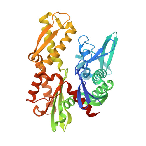
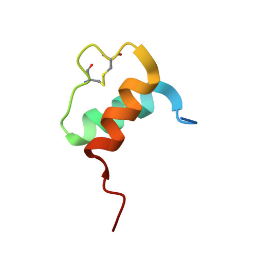
Crystal structure of the BiP NBD and MANF SAP complex


Experimental Data Snapshot

  • Method: X-RAY DIFFRACTION
  • Resolution: 1.57 Å
  • R-Value Free: 0.204 
  • R-Value Work: 0.184 
  • R-Value Observed: 0.185 

Starting Model: experimental
View more details

wwPDB Validation   3D Report Full Report


This is version 1.3 of the entry. See complete history


Literature

MANF antagonizes nucleotide exchange by the endoplasmic reticulum chaperone BiP.

Yan, Y.Rato, C.Rohland, L.Preissler, S.Ron, D.

(2019) Nat Commun 10: 541-541

  • DOI: https://doi.org/10.1038/s41467-019-08450-4
  • Primary Citation of Related Structures:  
    6H9U, 6HA7, 6HAB

  • PubMed Abstract: 

    Despite its known role as a secreted neuroprotectant, much of the mesencephalic astrocyte-derived neurotrophic factor (MANF) is retained in the endoplasmic reticulum (ER) of producer cells. There, by unknown mechanisms, MANF plays a role in protein folding homeostasis in complex with the ER-localized Hsp70 chaperone BiP. Here we report that the SAF-A/B, Acinus, and PIAS (SAP) domain of MANF selectively associates with the nucleotide binding domain (NBD) of ADP-bound BiP. In crystal structures the SAP domain engages the cleft between NBD subdomains Ia and IIa, stabilizing the ADP-bound conformation and clashing with the interdomain linker that occupies this site in ATP-bound BiP. MANF inhibits both ADP release from BiP and ATP binding to BiP, and thereby client release. Cells lacking MANF have fewer ER stress-induced BiP-containing high molecular weight complexes. These findings suggest that MANF contributes to protein folding homeostasis as a nucleotide exchange inhibitor that stabilizes certain BiP-client complexes.


  • Organizational Affiliation

    Cambridge Institute for Medical Research, University of Cambridge, Cambridge, CB2 0XY, UK.


Macromolecules
Find similar proteins by:  (by identity cutoff)  |  3D Structure
Entity ID: 1
MoleculeChains Sequence LengthOrganismDetailsImage
Endoplasmic reticulum chaperone BiP386Cricetulus griseusMutation(s): 0 
Gene Names: HSPA5GRP78I79_019946
EC: 3.6.4.10
UniProt
Find proteins for G3I8R9 (Cricetulus griseus)
Explore G3I8R9 
Go to UniProtKB:  G3I8R9
Entity Groups  
Sequence Clusters30% Identity50% Identity70% Identity90% Identity95% Identity100% Identity
UniProt GroupG3I8R9
Sequence Annotations
Expand
  • Reference Sequence
Find similar proteins by:  (by identity cutoff)  |  3D Structure
Entity ID: 2
MoleculeChains Sequence LengthOrganismDetailsImage
Mesencephalic astrocyte-derived neurotrophic factor44Mus musculusMutation(s): 0 
Gene Names: ManfArmet
UniProt
Find proteins for Q9CXI5 (Mus musculus)
Explore Q9CXI5 
Go to UniProtKB:  Q9CXI5
Entity Groups  
Sequence Clusters30% Identity50% Identity70% Identity90% Identity95% Identity100% Identity
UniProt GroupQ9CXI5
Sequence Annotations
Expand
  • Reference Sequence
Experimental Data & Validation

Experimental Data

  • Method: X-RAY DIFFRACTION
  • Resolution: 1.57 Å
  • R-Value Free: 0.204 
  • R-Value Work: 0.184 
  • R-Value Observed: 0.185 
  • Space Group: C 1 2 1
Unit Cell:
Length ( Å )Angle ( ˚ )
a = 153.31α = 90
b = 66.75β = 106.54
c = 44.3γ = 90
Software Package:
Software NamePurpose
REFMACrefinement
XDSdata reduction
Aimlessdata scaling
PHASERphasing

Structure Validation

View Full Validation Report


Entry History & Funding Information

Deposition Data


Funding OrganizationLocationGrant Number
Wellcome TrustUnited Kingdom200848/Z/16/Z
Wellcome TrustUnited Kingdom100140

Revision History  (Full details and data files)

  • Version 1.0: 2019-02-06
    Type: Initial release
  • Version 1.1: 2019-02-13
    Changes: Data collection, Database references
  • Version 1.2: 2024-01-17
    Changes: Data collection, Database references, Derived calculations, Refinement description
  • Version 1.3: 2024-11-06
    Changes: Structure summary