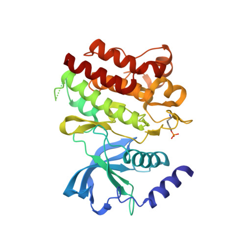
6I83

Crystal structure of phosphorylated RET V804M tyrosine kinase domain complexed with PDD00018366


Experimental Data Snapshot

  • Method: X-RAY DIFFRACTION
  • Resolution: 1.88 Å
  • R-Value Free: 0.238 
  • R-Value Work: 0.207 
  • R-Value Observed: 0.208 

Starting Model: experimental
View more details

wwPDB Validation   3D Report Full Report


Ligand Structure Quality Assessment 


This is version 1.3 of the entry. See complete history


Literature

Discovery and Optimization of wt-RET/KDR-Selective Inhibitors of RETV804MKinase.

Newton, R.Waszkowycz, B.Seewooruthun, C.Burschowsky, D.Richards, M.Hitchin, S.Begum, H.Watson, A.French, E.Hamilton, N.Jones, S.Lin, L.Y.Waddell, I.Echalier, A.Bayliss, R.Jordan, A.M.Ogilvie, D.

(2020) ACS Med Chem Lett 11: 497-505

  • DOI: https://doi.org/10.1021/acsmedchemlett.9b00615
  • Primary Citation of Related Structures:  
    6I82, 6I83

  • PubMed Abstract: 

    A combination of focused library and virtual screening, hit expansion, and rational design has resulted in the development of a series of inhibitors of RET V804M kinase, the anticipated drug-resistant mutant of RET kinase. These agents do not inhibit the wild type (wt) isoforms of RET or KDR and therefore offer a potential adjunct to RET inhibitors currently undergoing clinical evaluation.


  • Organizational Affiliation

    Drug Discovery Unit, Cancer Research UK, Manchester Institute, University of Manchester, Alderley Park, Macclesfield SK10 4TG, U.K.


Macromolecules
Find similar proteins by:  (by identity cutoff)  |  3D Structure
Entity ID: 1
MoleculeChains Sequence LengthOrganismDetailsImage
Proto-oncogene tyrosine-protein kinase receptor Ret314Homo sapiensMutation(s): 1 
Gene Names: RETCDHF12CDHR16PTCRET51
EC: 2.7.10.1
UniProt & NIH Common Fund Data Resources
Find proteins for P07949 (Homo sapiens)
Explore P07949 
Go to UniProtKB:  P07949
PHAROS:  P07949
GTEx:  ENSG00000165731 
Entity Groups  
Sequence Clusters30% Identity50% Identity70% Identity90% Identity95% Identity100% Identity
UniProt GroupP07949
Sequence Annotations
Expand
  • Reference Sequence
Small Molecules
Ligands 2 Unique
IDChains Name / Formula / InChI Key2D Diagram3D Interactions
H72
Query on H72

Download Ideal Coordinates CCD File 
B [auth A]4-[5-(pyridin-3-ylmethylamino)pyrazolo[1,5-a]pyrimidin-3-yl]benzamide
C19 H16 N6 O
LMGXQBQAUALFTB-UHFFFAOYSA-N
FMT
Query on FMT

Download Ideal Coordinates CCD File 
C [auth A]
D [auth A]
E [auth A]
F [auth A]
G [auth A]
C [auth A],
D [auth A],
E [auth A],
F [auth A],
G [auth A],
H [auth A],
I [auth A],
J [auth A],
K [auth A],
L [auth A],
M [auth A],
N [auth A],
O [auth A],
P [auth A],
Q [auth A]
FORMIC ACID
C H2 O2
BDAGIHXWWSANSR-UHFFFAOYSA-N
Modified Residues  1 Unique
IDChains TypeFormula2D DiagramParent
PTR
Query on PTR
A
L-PEPTIDE LINKINGC9 H12 N O6 PTYR
Binding Affinity Annotations 
IDSourceBinding Affinity
H72 BindingDB:  6I83 IC50: min: 51, max: 3900 (nM) from 2 assay(s)
Experimental Data & Validation

Experimental Data

  • Method: X-RAY DIFFRACTION
  • Resolution: 1.88 Å
  • R-Value Free: 0.238 
  • R-Value Work: 0.207 
  • R-Value Observed: 0.208 
  • Space Group: C 1 2 1
Unit Cell:
Length ( Å )Angle ( ˚ )
a = 72.38α = 90
b = 70.04β = 102.22
c = 78.73γ = 90
Software Package:
Software NamePurpose
GDAdata collection
XDSdata reduction
XSCALEdata scaling
PHASERphasing
REFMACrefinement

Structure Validation

View Full Validation Report


Ligand Structure Quality Assessment 


Entry History & Funding Information

Deposition Data


Funding OrganizationLocationGrant Number
Cancer Research UKUnited KingdomGrant C480/A11411
Cancer Research UKUnited KingdomC5759/A17098
Cancer Research UKUnited KingdomC1362/A20263

Revision History  (Full details and data files)

  • Version 1.0: 2020-03-11
    Type: Initial release
  • Version 1.1: 2020-04-22
    Changes: Database references
  • Version 1.2: 2024-01-24
    Changes: Data collection, Database references, Refinement description
  • Version 1.3: 2024-11-13
    Changes: Structure summary