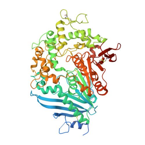
Decomposition of PET film by MHETase using Exo-PETase function
Sagong, H.-Y., Seo, H., Kim, T., Son, H., Joo, S., Lee, S., Kim, S., Woo, J.-S., Hwang, S., Kim, K.-J.(2020) ACS Catal 10: 4805-4812
Experimental Data Snapshot
(2020) ACS Catal 10: 4805-4812
Entity ID: 1 | |||||
|---|---|---|---|---|---|
| Molecule | Chains | Sequence Length | Organism | Details | Image |
| Mono(2-hydroxyethyl) terephthalate hydrolase | 621 | Piscinibacter sakaiensis | Mutation(s): 0 EC: 3.1.1.102 |  | |
UniProt | |||||
Find proteins for A0A0K8P8E7 (Piscinibacter sakaiensis) Explore A0A0K8P8E7 Go to UniProtKB: A0A0K8P8E7 | |||||
Entity Groups | |||||
| Sequence Clusters | 30% Identity50% Identity70% Identity90% Identity95% Identity100% Identity | ||||
| UniProt Group | A0A0K8P8E7 | ||||
Sequence AnnotationsExpand | |||||
| |||||
| Ligands 3 Unique | |||||
|---|---|---|---|---|---|
| ID | Chains | Name / Formula / InChI Key | 2D Diagram | 3D Interactions | |
| C8X Query on C8X | F [auth B] | bis(2-hydroxyethyl) benzene-1,4-dicarboxylate C12 H14 O6 QPKOBORKPHRBPS-UHFFFAOYSA-N |  | ||
| C9C Query on C9C | D [auth A], H [auth C] | 4-(2-hydroxyethyloxycarbonyl)benzoic acid C10 H10 O5 BCBHDSLDGBIFIX-UHFFFAOYSA-N |  | ||
| CA Query on CA | E [auth A], G [auth B], I [auth C] | CALCIUM ION Ca BHPQYMZQTOCNFJ-UHFFFAOYSA-N |  | ||
| Length ( Å ) | Angle ( ˚ ) |
|---|---|
| a = 215.259 | α = 90 |
| b = 173.799 | β = 119.51 |
| c = 106.671 | γ = 90 |
| Software Name | Purpose |
|---|---|
| REFMAC | refinement |
| PDB_EXTRACT | data extraction |
| HKL-2000 | data reduction |
| PHASER | phasing |
| HKL-2000 | data scaling |