6LQ6

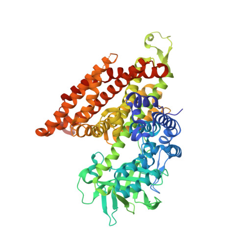
Crystal Structure of E447A Acyl-CoA Dehydrogenase FadE5 mutant from Mycobacteria smegmatis in complex with C20CoA


Experimental Data Snapshot

  • Method: X-RAY DIFFRACTION
  • Resolution: 2.00 Å
  • R-Value Free: 0.204 
  • R-Value Work: 0.162 
  • R-Value Observed: 0.164 

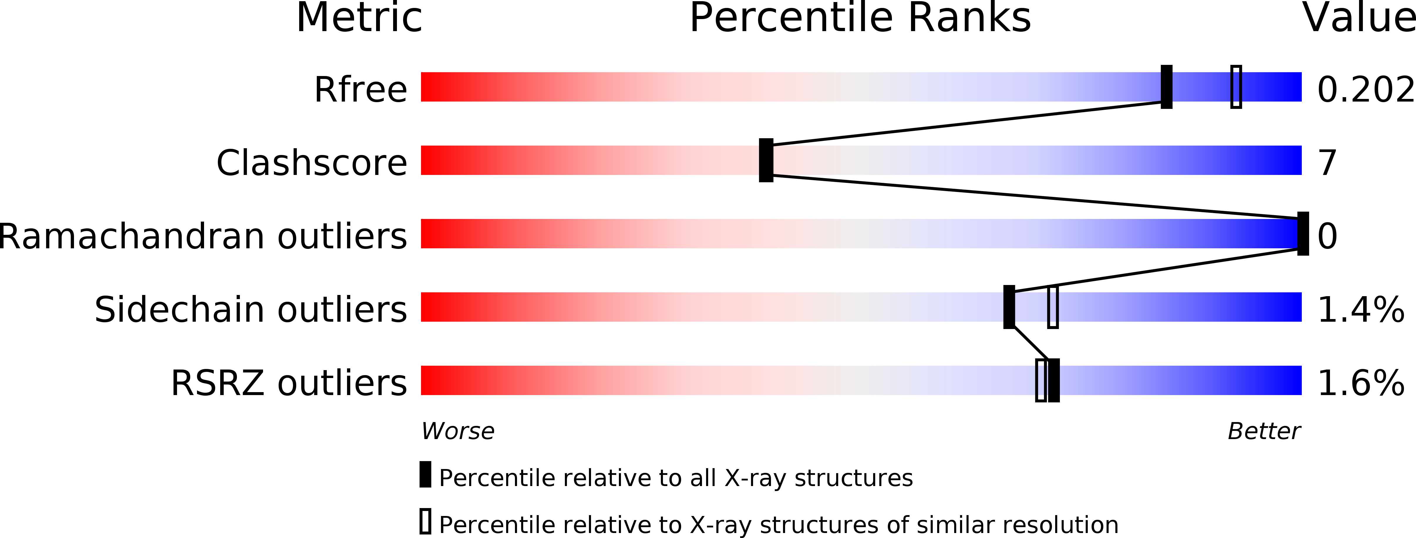
wwPDB Validation   3D Report Full Report


Ligand Structure Quality Assessment 


This is version 1.3 of the entry. See complete history


Literature

Structural basis for the broad substrate specificity of two acyl-CoA dehydrogenases FadE5 from mycobacteria.

Chen, X.Chen, J.Yan, B.Zhang, W.Guddat, L.W.Liu, X.Rao, Z.

(2020) Proc Natl Acad Sci U S A 117: 16324-16332

  • DOI: https://doi.org/10.1073/pnas.2002835117
  • Primary Citation of Related Structures:  
    6KPT, 6KRI, 6KS9, 6KSA, 6KSB, 6KSE, 6LPY, 6LQ0, 6LQ1, 6LQ2, 6LQ3, 6LQ4, 6LQ5, 6LQ6, 6LQ7, 6LQ8

  • PubMed Abstract: 

    FadE, an acyl-CoA dehydrogenase, introduces unsaturation to carbon chains in lipid metabolism pathways. Here, we report that FadE5 from Mycobacterium tuberculosis ( Mtb FadE5) and Mycobacterium smegmatis ( Ms FadE5) play roles in drug resistance and exhibit broad specificity for linear acyl-CoA substrates but have a preference for those with long carbon chains. Here, the structures of Ms FadE5 and Mtb FadE5, in the presence and absence of substrates, have been determined. These reveal the molecular basis for the broad substrate specificity of these enzymes. FadE5 interacts with the CoA region of the substrate through a large number of hydrogen bonds and an unusual π-π stacking interaction, allowing these enzymes to accept both short- and long-chain substrates. Residues in the substrate binding cavity reorient their side chains to accommodate substrates of various lengths. Longer carbon-chain substrates make more numerous hydrophobic interactions with the enzyme compared with the shorter-chain substrates, resulting in a preference for this type of substrate.


  • Organizational Affiliation

    State Key Laboratory of Medicinal Chemical Biology, Frontiers Science Center for Cell Responses, College of Life Sciences, Nankai University, 300071 Tianjin, China.


Macromolecules
Find similar proteins by:  (by identity cutoff)  |  3D Structure
Entity ID: 1
MoleculeChains Sequence LengthOrganismDetailsImage
Acyl-CoA dehydrogenase
A, B
612Mycolicibacterium smegmatisMutation(s): 1 
Gene Names: fadE5ERS451418_00380
EC: 1.3.8.1 (PDB Primary Data), 1.3.8.7 (UniProt), 1.3.8.8 (UniProt)
UniProt
Find proteins for Q3L887 (Mycolicibacterium smegmatis (strain ATCC 700084 / mc(2)155))
Explore Q3L887 
Go to UniProtKB:  Q3L887
Entity Groups  
Sequence Clusters30% Identity50% Identity70% Identity90% Identity95% Identity100% Identity
UniProt GroupQ3L887
Sequence Annotations
Expand
  • Reference Sequence
Experimental Data & Validation

Experimental Data

  • Method: X-RAY DIFFRACTION
  • Resolution: 2.00 Å
  • R-Value Free: 0.204 
  • R-Value Work: 0.162 
  • R-Value Observed: 0.164 
  • Space Group: P 21 21 2
Unit Cell:
Length ( Å )Angle ( ˚ )
a = 98.164α = 90
b = 206.572β = 90
c = 74.134γ = 90
Software Package:
Software NamePurpose
PHENIXrefinement
XSCALEdata scaling
PDB_EXTRACTdata extraction
XDSdata reduction
FFTphasing

Structure Validation

View Full Validation Report


Ligand Structure Quality Assessment 


Entry History 

Deposition Data

Revision History  (Full details and data files)

  • Version 1.0: 2020-07-01
    Type: Initial release
  • Version 1.1: 2020-07-15
    Changes: Database references
  • Version 1.2: 2020-07-29
    Changes: Database references
  • Version 1.3: 2024-03-27
    Changes: Data collection, Database references