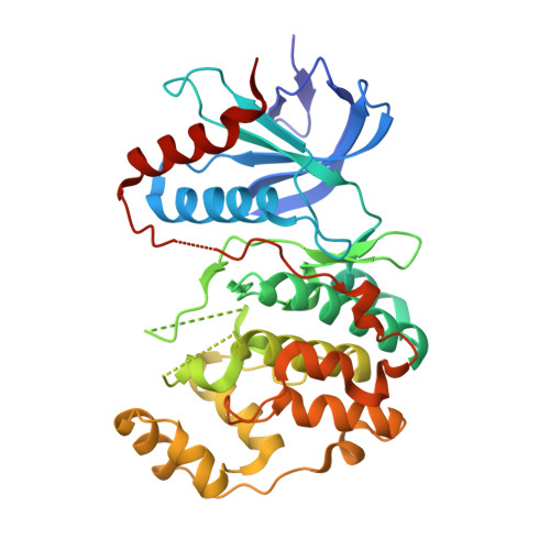
6OPI

phosphorylated ERK2 with SCH-CPD336

  • Classification: TRANSFERASE
  • Organism(s): Homo sapiens
  • Expression System: Escherichia coli
  • Mutation(s): No 

  • Deposited: 2019-04-25 Released: 2019-07-31 
  • Deposition Author(s): Vigers, G.P., Smith, D.
  • Funding Organization(s): National Institutes of Health/National Human Genome Research Institute (NIH/NHGRI)

Experimental Data Snapshot

  • Method: X-RAY DIFFRACTION
  • Resolution: 3.00 Å
  • R-Value Free: 0.260 
  • R-Value Work: 0.185 
  • R-Value Observed: 0.189 

Starting Model: experimental
View more details

wwPDB Validation   3D Report Full Report


Ligand Structure Quality Assessment 


This is version 1.5 of the entry. See complete history


Literature

Activation loop dynamics are controlled by conformation-selective inhibitors of ERK2.

Pegram, L.M.Liddle, J.C.Xiao, Y.Hoh, M.Rudolph, J.Iverson, D.B.Vigers, G.P.Smith, D.Zhang, H.Wang, W.Moffat, J.G.Ahn, N.G.

(2019) Proc Natl Acad Sci U S A 116: 15463-15468

  • DOI: https://doi.org/10.1073/pnas.1906824116
  • Primary Citation of Related Structures:  
    6OPG, 6OPH, 6OPI, 6OPK

  • PubMed Abstract: 

    Conformational selection by small molecules expands inhibitory possibilities for protein kinases. Nuclear magnetic resonance (NMR) measurements of the mitogen-activated protein (MAP) kinase ERK2 have shown that activation by dual phosphorylation induces global motions involving exchange between two states, L and R. We show that ERK inhibitors Vertex-11e and SCH772984 exploit the small energetic difference between L and R to shift the equilibrium in opposing directions. An X-ray structure of active 2P-ERK2 complexed with AMP-PNP reveals a shift in the Gly-rich loop along with domain closure to position the nucleotide in a more catalytically productive conformation relative to inactive 0P-ERK2:ATP. X-ray structures of 2P-ERK2 complexed with Vertex-11e or GDC-0994 recapitulate this closure, which is blocked in a complex with a SCH772984 analog. Thus, the L→R shift in 2P-ERK2 is associated with movements needed to form a competent active site. Solution measurements by hydrogen-exchange mass spectrometry (HX-MS) reveal distinct binding interactions for Vertex-11e, GDC-0994, and AMP-PNP with active vs. inactive ERK2, where the extent of HX protection correlates with R state formation. Furthermore, Vertex-11e and SCH772984 show opposite effects on HX near the activation loop. Consequently, these inhibitors differentially affect MAP kinase phosphatase activity toward 2P-ERK2. We conclude that global motions in ERK2 reflect conformational changes at the active site that promote productive nucleotide binding and couple with changes at the activation loop to allow control of dephosphorylation by conformationally selective inhibitors.


  • Organizational Affiliation

    Department of Biochemistry, University of Colorado, Boulder, CO 80305.


Macromolecules
Find similar proteins by:  (by identity cutoff)  |  3D Structure
Entity ID: 1
MoleculeChains Sequence LengthOrganismDetailsImage
Mitogen-activated protein kinase 1354Homo sapiensMutation(s): 0 
Gene Names: MAPK1ERK2PRKM1PRKM2
EC: 2.7.11.24
UniProt & NIH Common Fund Data Resources
Find proteins for P28482 (Homo sapiens)
Explore P28482 
Go to UniProtKB:  P28482
PHAROS:  P28482
GTEx:  ENSG00000100030 
Entity Groups  
Sequence Clusters30% Identity50% Identity70% Identity90% Identity95% Identity100% Identity
UniProt GroupP28482
Sequence Annotations
Expand
  • Reference Sequence
Small Molecules
Ligands 1 Unique
IDChains Name / Formula / InChI Key2D Diagram3D Interactions
N0V (Subject of Investigation/LOI)
Query on N0V

Download Ideal Coordinates CCD File 
B [auth A](3R)-N-[3-(2-cyclopropylpyridin-4-yl)-1H-indazol-5-yl]-3-(methoxymethyl)-1-(2-oxo-2-{4-[4-(pyrimidin-2-yl)phenyl]-3,6-dihydropyridin-1(2H)-yl}ethyl)pyrrolidine-3-carboxamide
C39 H40 N8 O3
FSTCYTJKYVEAES-LDLOPFEMSA-N
Modified Residues  2 Unique
IDChains TypeFormula2D DiagramParent
PTR
Query on PTR
A
L-PEPTIDE LINKINGC9 H12 N O6 PTYR
TPO
Query on TPO
A
L-PEPTIDE LINKINGC4 H10 N O6 PTHR
Experimental Data & Validation

Experimental Data

  • Method: X-RAY DIFFRACTION
  • Resolution: 3.00 Å
  • R-Value Free: 0.260 
  • R-Value Work: 0.185 
  • R-Value Observed: 0.189 
  • Space Group: P 21 21 21
Unit Cell:
Length ( Å )Angle ( ˚ )
a = 41.394α = 90
b = 77.64β = 90
c = 126.536γ = 90
Software Package:
Software NamePurpose
REFMACrefinement
MOSFLMdata reduction
Aimlessdata scaling
PHASERphasing

Structure Validation

View Full Validation Report


Ligand Structure Quality Assessment 


Entry History & Funding Information

Deposition Data


Funding OrganizationLocationGrant Number
National Institutes of Health/National Human Genome Research Institute (NIH/NHGRI)United StatesR01GM114594
National Institutes of Health/National Human Genome Research Institute (NIH/NHGRI)United StatesS10RR026641
National Institutes of Health/National Human Genome Research Institute (NIH/NHGRI)United StatesT32GM008759

Revision History  (Full details and data files)

  • Version 1.0: 2019-07-31
    Type: Initial release
  • Version 1.1: 2019-08-14
    Changes: Data collection, Database references
  • Version 1.2: 2019-12-18
    Changes: Author supporting evidence
  • Version 1.3: 2023-10-11
    Changes: Data collection, Database references, Refinement description
  • Version 1.4: 2023-11-15
    Changes: Data collection
  • Version 1.5: 2024-10-23
    Changes: Structure summary