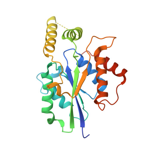
6U03

Crystal Structure of Fungal RNA Kinase


Experimental Data Snapshot

  • Method: X-RAY DIFFRACTION
  • Resolution: 1.85 Å
  • R-Value Free: 0.215 
  • R-Value Work: 0.170 
  • R-Value Observed: 0.174 

Starting Model: experimental
View more details

wwPDB Validation   3D Report Full Report


Ligand Structure Quality Assessment 


This is version 1.4 of the entry. See complete history


Literature

Atomic structures of the RNA end-healing 5'-OH kinase and 2',3'-cyclic phosphodiesterase domains of fungal tRNA ligase: conformational switches in the kinase upon binding of the GTP phosphate donor.

Banerjee, A.Goldgur, Y.Schwer, B.Shuman, S.

(2019) Nucleic Acids Res 47: 11826-11838

  • DOI: https://doi.org/10.1093/nar/gkz1049
  • Primary Citation of Related Structures:  
    6TZM, 6TZO, 6TZX, 6U00, 6U03, 6U05

  • PubMed Abstract: 

    Fungal tRNA ligase (Trl1) rectifies RNA breaks with 2',3'-cyclic-PO4 and 5'-OH termini. Trl1 consists of three catalytic modules: an N-terminal ligase (LIG) domain; a central polynucleotide kinase (KIN) domain; and a C-terminal cyclic phosphodiesterase (CPD) domain. Trl1 enzymes found in all human fungal pathogens are untapped targets for antifungal drug discovery. Here we report a 1.9 Å crystal structure of Trl1 KIN-CPD from the pathogenic fungus Candida albicans, which adopts an extended conformation in which separate KIN and CPD domains are connected by an unstructured linker. CPD belongs to the 2H phosphotransferase superfamily by dint of its conserved central concave β sheet and interactions of its dual HxT motif histidines and threonines with phosphate in the active site. Additional active site motifs conserved among the fungal CPD clade of 2H enzymes are identified. We present structures of the Candida Trl1 KIN domain at 1.5 to 2.0 Å resolution-as apoenzyme and in complexes with GTP•Mg2+, IDP•PO4, and dGDP•PO4-that highlight conformational switches in the G-loop (which recognizes the guanine base) and lid-loop (poised over the nucleotide phosphates) that accompany nucleotide binding.


  • Organizational Affiliation

    Molecular Biology Program, Sloan-Kettering Institute, New York, NY 10065, USA.


Macromolecules
Find similar proteins by:  (by identity cutoff)  |  3D Structure
Entity ID: 1
MoleculeChains Sequence LengthOrganismDetailsImage
tRNA ligase235Candida albicans SC5314Mutation(s): 1 
Gene Names: LIG1RLG1TRL1CAALFM_C702060WACaJ7.0238CaO19.13864CaO19.6511
EC: 6.5.1.3
UniProt
Find proteins for P43075 (Candida albicans (strain SC5314 / ATCC MYA-2876))
Explore P43075 
Go to UniProtKB:  P43075
Entity Groups  
Sequence Clusters30% Identity50% Identity70% Identity90% Identity95% Identity100% Identity
UniProt GroupP43075
Sequence Annotations
Expand
  • Reference Sequence
Experimental Data & Validation

Experimental Data

  • Method: X-RAY DIFFRACTION
  • Resolution: 1.85 Å
  • R-Value Free: 0.215 
  • R-Value Work: 0.170 
  • R-Value Observed: 0.174 
  • Space Group: P 21 21 21
Unit Cell:
Length ( Å )Angle ( ˚ )
a = 50.227α = 90
b = 55.667β = 90
c = 81.795γ = 90
Software Package:
Software NamePurpose
PHENIXrefinement
HKL-2000data reduction
HKL-2000data scaling
PHENIXphasing

Structure Validation

View Full Validation Report


Ligand Structure Quality Assessment 


Entry History & Funding Information

Deposition Data


Funding OrganizationLocationGrant Number
National Institutes of Health/National Institute of General Medical Sciences (NIH/NIGMS)United StatesGM42498

Revision History  (Full details and data files)

  • Version 1.0: 2019-11-06
    Type: Initial release
  • Version 1.1: 2019-11-27
    Changes: Database references
  • Version 1.2: 2019-12-25
    Changes: Database references
  • Version 1.3: 2020-01-01
    Changes: Author supporting evidence
  • Version 1.4: 2023-10-11
    Changes: Data collection, Database references, Derived calculations, Refinement description