6WF7

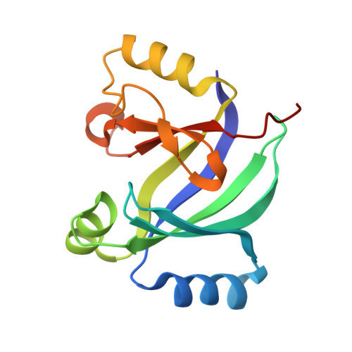
Methylmalonyl-CoA epimerase in complex with methylmalonyl-CoA and NH4+


Experimental Data Snapshot

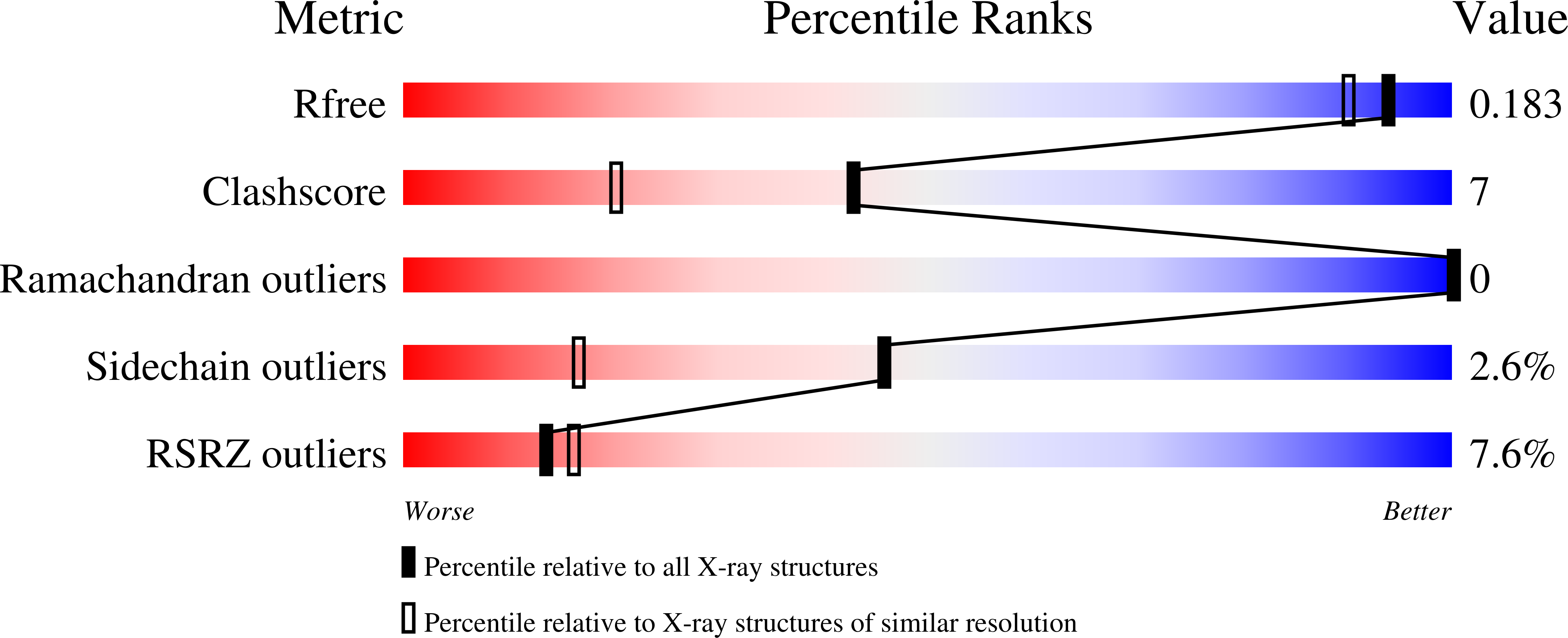
  • Method: X-RAY DIFFRACTION
  • Resolution: 1.55 Å
  • R-Value Free: 0.170 
  • R-Value Work: 0.151 
  • R-Value Observed: 0.152 

Starting Model: experimental
View more details

wwPDB Validation   3D Report Full Report


Ligand Structure Quality Assessment 


This is version 1.3 of the entry. See complete history


Literature

Substrate Enolate Intermediate and Mimic Captured in the Active Site of Streptomyces coelicolor Methylmalonyl-CoA Epimerase.

Stunkard, L.M.Benjamin, A.B.Bower, J.B.Huth, T.J.Lohman, J.R.

(2022) Chembiochem 23: e202100487-e202100487

  • DOI: https://doi.org/10.1002/cbic.202100487
  • Primary Citation of Related Structures:  
    6WF6, 6WF7, 6WFH, 6WFI

  • PubMed Abstract: 

    Methylmalonyl-CoA epimerase (MMCE) is proposed to use general acid-base catalysis, but the proposed catalytic glutamic acids are highly asymmetrical in the active site unlike many other racemases. To gain insight into the puzzling relationships between catalytic mechanism, structure, and substrate preference, we solved Streptomyces coelicolor MMCE structures with substrate or 2-nitropropionyl-CoA, an intermediate/transition state analogue. Both ligand bound structures have a planar methylmalonate/2-nitropropionyl moiety indicating a deprotonated C2 with ≥4 Å distances to either catalytic acid. Both glutamates interact with the carboxylate/nitro group, either directly or through other residues. This suggests the proposed catalytic acids sequentially catalyze proton shifts between C2 and carboxylate of the substrate with an enolate intermediate. In addition, our structures provide a platform to design mutations for expanding substrate scope to support combinatorial biosynthesis.


  • Organizational Affiliation

    Department of Biochemistry, Purdue University, 175 S. University St., West Lafayette, IN 47907, USA.


Macromolecules
Find similar proteins by:  (by identity cutoff)  |  3D Structure
Entity ID: 1
MoleculeChains Sequence LengthOrganismDetailsImage
Methylmalonyl-CoA epimerase146Streptomyces coelicolor A3(2)Mutation(s): 1 
Gene Names: SCO5398
UniProt
Find proteins for Q9L2C2 (Streptomyces coelicolor (strain ATCC BAA-471 / A3(2) / M145))
Explore Q9L2C2 
Go to UniProtKB:  Q9L2C2
Entity Groups  
Sequence Clusters30% Identity50% Identity70% Identity90% Identity95% Identity100% Identity
UniProt GroupQ9L2C2
Sequence Annotations
Expand
  • Reference Sequence
Small Molecules
Ligands 4 Unique
IDChains Name / Formula / InChI Key2D Diagram3D Interactions
MC0 (Subject of Investigation/LOI)
Query on MC0

Download Ideal Coordinates CCD File 
E [auth A](S)-Methylmalonyl-Coenzyme A
C25 H40 N7 O19 P3 S
MZFOKIKEPGUZEN-IBNUZSNCSA-N
MCA (Subject of Investigation/LOI)
Query on MCA

Download Ideal Coordinates CCD File 
F [auth A]METHYLMALONYL-COENZYME A
C25 H40 N7 O19 P3 S
MZFOKIKEPGUZEN-AGCMQPJKSA-N
SO4
Query on SO4

Download Ideal Coordinates CCD File 
C [auth A],
D [auth A]
SULFATE ION
O4 S
QAOWNCQODCNURD-UHFFFAOYSA-L
NH4
Query on NH4

Download Ideal Coordinates CCD File 
B [auth A]AMMONIUM ION
H4 N
QGZKDVFQNNGYKY-UHFFFAOYSA-O
Experimental Data & Validation

Experimental Data

  • Method: X-RAY DIFFRACTION
  • Resolution: 1.55 Å
  • R-Value Free: 0.170 
  • R-Value Work: 0.151 
  • R-Value Observed: 0.152 
  • Space Group: P 41 21 2
Unit Cell:
Length ( Å )Angle ( ˚ )
a = 68.537α = 90
b = 68.537β = 90
c = 103.15γ = 90
Software Package:
Software NamePurpose
REFMACrefinement
HKL-2000data scaling
PDB_EXTRACTdata extraction
ARP/wARPmodel building
PHASERphasing
HKL-2000data reduction

Structure Validation

View Full Validation Report


Ligand Structure Quality Assessment 


Entry History 

Deposition Data

Revision History  (Full details and data files)

  • Version 1.0: 2020-07-08
    Type: Initial release
  • Version 1.1: 2022-01-12
    Changes: Database references, Source and taxonomy
  • Version 1.2: 2022-01-26
    Changes: Database references
  • Version 1.3: 2023-10-18
    Changes: Data collection, Refinement description