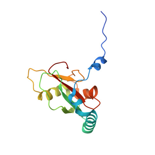
7BRQ

Crystal structure of human FAM134B LIR fused to human GABARAP


Experimental Data Snapshot

  • Method: X-RAY DIFFRACTION
  • Resolution: 1.40 Å
  • R-Value Free: 0.206 
  • R-Value Work: 0.192 
  • R-Value Observed: 0.192 

Starting Model: experimental
View more details

wwPDB Validation   3D Report Full Report


This is version 1.2 of the entry. See complete history


Literature

Super-assembly of ER-phagy receptor Atg40 induces local ER remodeling at contacts with forming autophagosomal membranes.

Mochida, K.Yamasaki, A.Matoba, K.Kirisako, H.Noda, N.N.Nakatogawa, H.

(2020) Nat Commun 11: 3306-3306

  • DOI: https://doi.org/10.1038/s41467-020-17163-y
  • Primary Citation of Related Structures:  
    7BRN, 7BRQ, 7BRT, 7BRU

  • PubMed Abstract: 

    The endoplasmic reticulum (ER) is selectively degraded by autophagy (ER-phagy) through proteins called ER-phagy receptors. In Saccharomyces cerevisiae, Atg40 acts as an ER-phagy receptor to sequester ER fragments into autophagosomes by binding Atg8 on forming autophagosomal membranes. During ER-phagy, parts of the ER are morphologically rearranged, fragmented, and loaded into autophagosomes, but the mechanism remains poorly understood. Here we find that Atg40 molecules assemble in the ER membrane concurrently with autophagosome formation via multivalent interaction with Atg8. Atg8-mediated super-assembly of Atg40 generates highly-curved ER regions, depending on its reticulon-like domain, and supports packing of these regions into autophagosomes. Moreover, tight binding of Atg40 to Atg8 is achieved by a short helix C-terminal to the Atg8-family interacting motif, and this feature is also observed for mammalian ER-phagy receptors. Thus, this study significantly advances our understanding of the mechanisms of ER-phagy and also provides insights into organelle fragmentation in selective autophagy of other organelles.


  • Organizational Affiliation

    School of Life Science and Technology, Tokyo Institute of Technology, Yokohama, Japan.


Macromolecules
Find similar proteins by:  (by identity cutoff)  |  3D Structure
Entity ID: 1
MoleculeChains Sequence LengthOrganismDetailsImage
Reticulophagy regulator 1,Gamma-aminobutyric acid receptor-associated protein137Homo sapiensMutation(s): 2 
Gene Names: FAM134BGABARAP
UniProt & NIH Common Fund Data Resources
Find proteins for Q9H6L5 (Homo sapiens)
Explore Q9H6L5 
Go to UniProtKB:  Q9H6L5
PHAROS:  Q9H6L5
GTEx:  ENSG00000154153 
Find proteins for O95166 (Homo sapiens)
Explore O95166 
Go to UniProtKB:  O95166
PHAROS:  O95166
GTEx:  ENSG00000170296 
Entity Groups  
Sequence Clusters30% Identity50% Identity70% Identity90% Identity95% Identity100% Identity
UniProt GroupsO95166Q9H6L5
Sequence Annotations
Expand
  • Reference Sequence
Small Molecules
Ligands 1 Unique
IDChains Name / Formula / InChI Key2D Diagram3D Interactions
GOL
Query on GOL

Download Ideal Coordinates CCD File 
B [auth A]GLYCEROL
C3 H8 O3
PEDCQBHIVMGVHV-UHFFFAOYSA-N
Experimental Data & Validation

Experimental Data

  • Method: X-RAY DIFFRACTION
  • Resolution: 1.40 Å
  • R-Value Free: 0.206 
  • R-Value Work: 0.192 
  • R-Value Observed: 0.192 
  • Space Group: P 63
Unit Cell:
Length ( Å )Angle ( ˚ )
a = 81.892α = 90
b = 81.892β = 90
c = 33.324γ = 120
Software Package:
Software NamePurpose
PHENIXrefinement
PDB_EXTRACTdata extraction
XDSdata reduction
XDSdata scaling
PHENIXphasing

Structure Validation

View Full Validation Report


Entry History & Funding Information

Deposition Data


Funding OrganizationLocationGrant Number
Japan Society for the Promotion of Science (JSPS)Japan17K18339
Japan Society for the Promotion of Science (JSPS)Japan19H05707
Japan Science and TechnologyJapanJPMJCR13M7

Revision History  (Full details and data files)

  • Version 1.0: 2020-07-08
    Type: Initial release
  • Version 1.1: 2020-07-22
    Changes: Database references
  • Version 1.2: 2023-11-29
    Changes: Data collection, Database references, Refinement description