RCSB PDB - 7FQP: PanDDA analysis group deposition -- Crystal structure of PTP1B in complex with FMOOA000505a

 7FQP

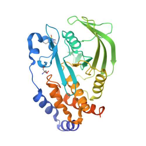
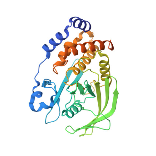
PanDDA analysis group deposition -- Crystal structure of PTP1B in complex with FMOOA000505a


Experimental Data Snapshot

  • Method: X-RAY DIFFRACTION
  • Resolution: 1.88 Å
  • R-Value Free: 0.207 
  • R-Value Work: 0.177 
  • R-Value Observed: 0.178 

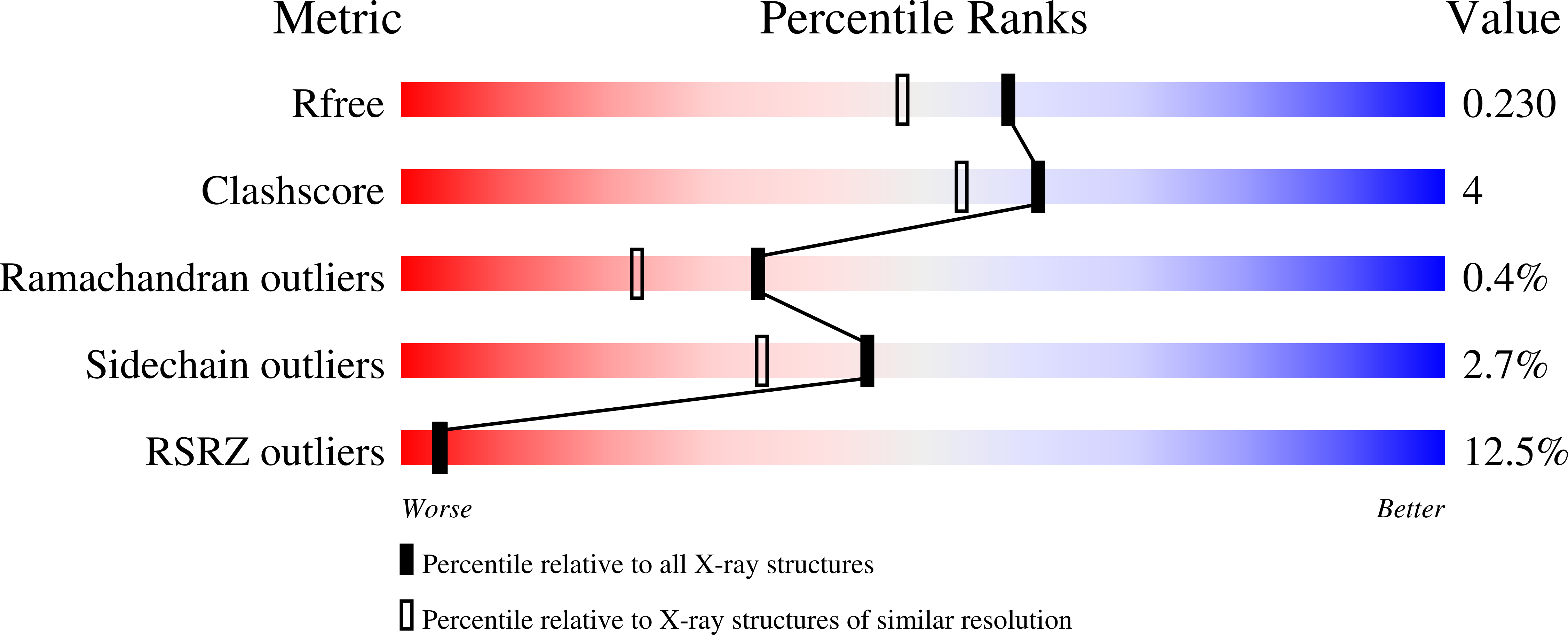
wwPDB Validation   3D Report Full Report


Ligand Structure Quality Assessment 

Created with Raphaël 2.3.0Worse 01 BetterLigand structure goodness of fit to experimental dataBest fitted JN1Click on this verticalbar to view details

This is version 1.2 of the entry. See complete history


Literature

Room-temperature crystallography reveals altered binding of small-molecule fragments to PTP1B.

Skaist Mehlman, T.Biel, J.T.Azeem, S.M.Nelson, E.R.Hossain, S.Dunnett, L.Paterson, N.G.Douangamath, A.Talon, R.Axford, D.Orins, H.von Delft, F.Keedy, D.A.

(2023) Elife 12

  • DOI: https://doi.org/10.7554/eLife.84632
  • Primary Citation of Related Structures:  
    7FQM, 7FQN, 7FQO, 7FQP, 7FQQ, 7FQR, 7FQS, 7FQT, 7FQU, 7FQV, 7FQW, 7FQX, 7FQY, 7FQZ, 7FRE, 7FRF, 7FRG, 7FRH, 7FRI, 7FRJ, 7FRK, 7FRL, 7FRM, 7FRN, 7FRO, 7FRP, 7FRQ, 7FRR, 7FRS, 7FRT, 7FRU

  • PubMed Abstract: 

    Much of our current understanding of how small-molecule ligands interact with proteins stems from X-ray crystal structures determined at cryogenic (cryo) temperature. For proteins alone, room-temperature (RT) crystallography can reveal previously hidden, biologically relevant alternate conformations. However, less is understood about how RT crystallography may impact the conformational landscapes of protein-ligand complexes. Previously, we showed that small-molecule fragments cluster in putative allosteric sites using a cryo crystallographic screen of the therapeutic target PTP1B (Keedy et al., 2018). Here, we have performed two RT crystallographic screens of PTP1B using many of the same fragments, representing the largest RT crystallographic screens of a diverse library of ligands to date, and enabling a direct interrogation of the effect of data collection temperature on protein-ligand interactions. We show that at RT, fewer ligands bind, and often more weakly - but with a variety of temperature-dependent differences, including unique binding poses, changes in solvation, new binding sites, and distinct protein allosteric conformational responses. Overall, this work suggests that the vast body of existing cryo-temperature protein-ligand structures may provide an incomplete picture, and highlights the potential of RT crystallography to help complete this picture by revealing distinct conformational modes of protein-ligand systems. Our results may inspire future use of RT crystallography to interrogate the roles of protein-ligand conformational ensembles in biological function.


  • Organizational Affiliation

    Structural Biology Initiative, CUNY Advanced Science Research Center, New York, United States.


Macromolecules
Find similar proteins by:  (by identity cutoff)  |  3D Structure
Entity ID: 1
MoleculeChains Sequence LengthOrganismDetailsImage
Tyrosine-protein phosphatase non-receptor type 1321Homo sapiensMutation(s): 2 
Gene Names: PTPN1PTP1B
EC: 3.1.3.48
UniProt & NIH Common Fund Data Resources
Find proteins for P18031 (Homo sapiens)
Explore P18031 
Go to UniProtKB:  P18031
PHAROS:  P18031
GTEx:  ENSG00000196396 
Entity Groups  
Sequence Clusters30% Identity50% Identity70% Identity90% Identity95% Identity100% Identity
UniProt GroupP18031
Sequence Annotations
Expand
  • Reference Sequence
Small Molecules
Ligands 2 Unique
IDChains Name / Formula / InChI Key2D Diagram3D Interactions
JN1 (Subject of Investigation/LOI)
Query on JN1

Download Ideal Coordinates CCD File 
C [auth A](6R)-5,6-dihydro-1H-2,6-methano-1lambda~6~-1lambda~6~,2,5-benzothiadiazocine-1,1,4(3H)-trione
C10 H10 N2 O3 S
OILUHIZMKUPPSN-QMMMGPOBSA-N
TRS
Query on TRS

Download Ideal Coordinates CCD File 
B [auth A]2-AMINO-2-HYDROXYMETHYL-PROPANE-1,3-DIOL
C4 H12 N O3
LENZDBCJOHFCAS-UHFFFAOYSA-O
Experimental Data & Validation

Experimental Data

  • Method: X-RAY DIFFRACTION
  • Resolution: 1.88 Å
  • R-Value Free: 0.207 
  • R-Value Work: 0.177 
  • R-Value Observed: 0.178 
  • Space Group: P 31 2 1
Unit Cell:
Length ( Å )Angle ( ˚ )
a = 89.56α = 90
b = 89.56β = 90
c = 106.38γ = 120
Software Package:
Software NamePurpose
REFMACrefinement
XSCALEdata scaling
PDB_EXTRACTdata extraction
XDSdata reduction
DIMPLEphasing

Structure Validation

View Full Validation Report


Ligand Structure Quality Assessment 

Created with Raphaël 2.3.0Worse 01 BetterLigand structure goodness of fit to experimental dataBest fitted JN1Click on this verticalbar to view details

Entry History & Funding Information

Deposition Data


Funding OrganizationLocationGrant Number
U.S. Department of Education Graduate Assistance in Areas of National Need (GAANN)United StatesPA200A150068
National Institutes of Health/National Institute of General Medical Sciences (NIH/NIGMS)United StatesGM133769

Revision History  (Full details and data files)

  • Version 1.0: 2022-11-23
    Type: Initial release
  • Version 1.1: 2023-03-22
    Changes: Database references
  • Version 1.2: 2024-05-22
    Changes: Data collection