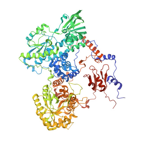
7M32

Dihydropyrimidine Dehydrogenase (DPD) C671A Mutant Soaked with Uracil and NADPH Anaerobically


Experimental Data Snapshot

  • Method: X-RAY DIFFRACTION
  • Resolution: 1.82 Å
  • R-Value Free: 0.202 
  • R-Value Work: 0.169 
  • R-Value Observed: 0.171 

Starting Model: experimental
View more details

wwPDB Validation   3D Report Full Report


Ligand Structure Quality Assessment 


This is version 1.2 of the entry. See complete history


Literature

Perturbing the Movement of Hydrogens to Delineate and Assign Events in the Reductive Activation and Turnover of Porcine Dihydropyrimidine Dehydrogenase.

Beaupre, B.A.Forouzesh, D.C.Butrin, A.Liu, D.Moran, G.R.

(2021) Biochemistry 60: 1764-1775

  • DOI: https://doi.org/10.1021/acs.biochem.1c00243
  • Primary Citation of Related Structures:  
    7M31, 7M32

  • PubMed Abstract: 

    The native function of dihydropyrimidine dehydrogenase (DPD) is to reduce the 5,6-vinylic bond of pyrimidines uracil and thymine with electrons obtained from NADPH. NADPH and pyrimidines bind at separate active sites separated by ∼60 Å that are bridged by four Fe 4 S 4 centers. We have shown that DPD undergoes reductive activation, taking up two electrons from NADPH [Beaupre, B. A., et al. (2020) Biochemistry 59 , 2419-2431]. pH studies indicate that the rate of turnover is not controlled by the protonation state of the general acid, cysteine 671. The activation of the C671 variants is delineated into two phases particularly at low pH values. Spectral deconvolution of the delineated reductive activation reaction reveals that the initial phase results in the accumulation of charge transfer absorption added to the binding difference spectrum for NADPH. The second phase results in reduction of one of the two flavins. X-ray crystal structure analysis of the C671S variant soaked with NADPH and the slow substrate, thymine, in a low-oxygen atmosphere resolved the presumed activated form of the enzyme that has the FMN cofactor reduced. These data reveal that charge transfer arises from the proximity of the NADPH and FAD bases and that the ensuing flavin is a result of rapid transfer of electrons to the FMN without accumulation of reduced forms of the FAD or Fe 4 S 4 centers. These data suggest that the slow rate of turnover of DPD is governed by the movement of a mobile structural feature that carries the C671 residue.


  • Organizational Affiliation

    Department of Chemistry and Biochemistry, Loyola University Chicago, 1068 West Sheridan Road, Chicago, Illinois 60660, United States.


Macromolecules
Find similar proteins by:  (by identity cutoff)  |  3D Structure
Entity ID: 1
MoleculeChains Sequence LengthOrganismDetailsImage
Dihydropyrimidine dehydrogenase [NADP(+)]
A, B, C, D
1,025Sus scrofaMutation(s): 1 
Gene Names: DPYD
EC: 1.3.1.2
UniProt
Find proteins for Q28943 (Sus scrofa)
Explore Q28943 
Go to UniProtKB:  Q28943
Entity Groups  
Sequence Clusters30% Identity50% Identity70% Identity90% Identity95% Identity100% Identity
UniProt GroupQ28943
Sequence Annotations
Expand
  • Reference Sequence
Small Molecules
Ligands 7 Unique
IDChains Name / Formula / InChI Key2D Diagram3D Interactions
FAD (Subject of Investigation/LOI)
Query on FAD

Download Ideal Coordinates CCD File 
I [auth A],
IA [auth D],
R [auth B],
Z [auth C]
FLAVIN-ADENINE DINUCLEOTIDE
C27 H33 N9 O15 P2
VWWQXMAJTJZDQX-UYBVJOGSSA-N
FNR (Subject of Investigation/LOI)
Query on FNR

Download Ideal Coordinates CCD File 
BA [auth C],
K [auth A],
KA [auth D],
T [auth B]
1-DEOXY-1-(7,8-DIMETHYL-2,4-DIOXO-3,4-DIHYDRO-2H-BENZO[G]PTERIDIN-1-ID-10(5H)-YL)-5-O-PHOSPHONATO-D-RIBITOL
C17 H23 N4 O9 P
YTNIXZGTHTVJBW-SCRDCRAPSA-N
SF4 (Subject of Investigation/LOI)
Query on SF4

Download Ideal Coordinates CCD File 
E [auth A]
EA [auth D]
F [auth A]
FA [auth D]
G [auth A]
E [auth A],
EA [auth D],
F [auth A],
FA [auth D],
G [auth A],
GA [auth D],
H [auth A],
HA [auth D],
N [auth B],
O [auth B],
P [auth B],
Q [auth B],
V [auth C],
W [auth C],
X [auth C],
Y [auth C]
IRON/SULFUR CLUSTER
Fe4 S4
LJBDFODJNLIPKO-UHFFFAOYSA-N
LEU (Subject of Investigation/LOI)
Query on LEU

Download Ideal Coordinates CCD File 
CA [auth D],
M [auth B]
LEUCINE
C6 H13 N O2
ROHFNLRQFUQHCH-YFKPBYRVSA-N
PRO (Subject of Investigation/LOI)
Query on PRO

Download Ideal Coordinates CCD File 
U [auth C]PROLINE
C5 H9 N O2
ONIBWKKTOPOVIA-BYPYZUCNSA-N
URA (Subject of Investigation/LOI)
Query on URA

Download Ideal Coordinates CCD File 
AA [auth C],
J [auth A],
JA [auth D],
S [auth B]
URACIL
C4 H4 N2 O2
ISAKRJDGNUQOIC-UHFFFAOYSA-N
ALA (Subject of Investigation/LOI)
Query on ALA

Download Ideal Coordinates CCD File 
DA [auth D],
L [auth A]
ALANINE
C3 H7 N O2
QNAYBMKLOCPYGJ-REOHCLBHSA-N
Experimental Data & Validation

Experimental Data

  • Method: X-RAY DIFFRACTION
  • Resolution: 1.82 Å
  • R-Value Free: 0.202 
  • R-Value Work: 0.169 
  • R-Value Observed: 0.171 
  • Space Group: P 1 21 1
Unit Cell:
Length ( Å )Angle ( ˚ )
a = 81.87α = 90
b = 158.73β = 95.89
c = 162.7γ = 90
Software Package:
Software NamePurpose
Aimlessdata scaling
PHENIXrefinement
PDB_EXTRACTdata extraction
autoPROCdata reduction
PHASERphasing

Structure Validation

View Full Validation Report


Ligand Structure Quality Assessment 


Entry History 

Deposition Data

Revision History  (Full details and data files)

  • Version 1.0: 2021-06-09
    Type: Initial release
  • Version 1.1: 2021-06-23
    Changes: Database references
  • Version 1.2: 2023-10-18
    Changes: Advisory, Data collection, Database references, Refinement description