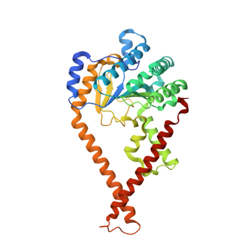
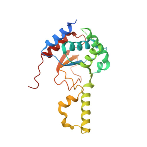
7PAY

Structure of the human heterotetrameric cis-prenyltransferase complex in complex with magnesium and GGsPP


Experimental Data Snapshot

  • Method: X-RAY DIFFRACTION
  • Resolution: 2.40 Å
  • R-Value Free: 0.244 
  • R-Value Work: 0.202 
  • R-Value Observed: 0.205 

Starting Model: experimental
View more details

wwPDB Validation   3D Report Full Report


Ligand Structure Quality Assessment 


This is version 1.1 of the entry. See complete history


Literature

Structural basis for long-chain isoprenoid synthesis by cis -prenyltransferases.

Giladi, M.Lisnyansky Bar-El, M.Vankova, P.Ferofontov, A.Melvin, E.Alkaderi, S.Kavan, D.Redko, B.Haimov, E.Wiener, R.Man, P.Haitin, Y.

(2022) Sci Adv 8: eabn1171-eabn1171

  • DOI: https://doi.org/10.1126/sciadv.abn1171
  • Primary Citation of Related Structures:  
    7PAX, 7PAY, 7PB0, 7PB1

  • PubMed Abstract: 

    Isoprenoids are synthesized by the prenyltransferase superfamily, which is subdivided according to the product stereoisomerism and length. In short- and medium-chain isoprenoids, product length correlates with active site volume. However, enzymes synthesizing long-chain products and rubber synthases fail to conform to this paradigm, because of an unexpectedly small active site. Here, we focused on the human cis -prenyltransferase complex (h cis -PT), residing at the endoplasmic reticulum membrane and playing a crucial role in protein glycosylation. Crystallographic investigation of h cis -PT along the reaction cycle revealed an outlet for the elongating product. Hydrogen-deuterium exchange mass spectrometry analysis showed that the hydrophobic active site core is flanked by dynamic regions consistent with separate inlet and outlet orifices. Last, using a fluorescence substrate analog, we show that product elongation and membrane association are closely correlated. Together, our results support direct membrane insertion of the elongating isoprenoid during catalysis, uncoupling active site volume from product length.


  • Organizational Affiliation

    Department of Physiology and Pharmacology, Sackler School of Medicine, Tel-Aviv University, Tel-Aviv 6997801, Israel.


Macromolecules
Find similar proteins by:  (by identity cutoff)  |  3D Structure
Entity ID: 1
MoleculeChains Sequence LengthOrganismDetailsImage
Dehydrodolichyl diphosphate synthase complex subunit DHDDS340Homo sapiensMutation(s): 0 
Gene Names: DHDDSHDS
EC: 2.5.1.87
UniProt & NIH Common Fund Data Resources
Find proteins for Q86SQ9 (Homo sapiens)
Explore Q86SQ9 
Go to UniProtKB:  Q86SQ9
PHAROS:  Q86SQ9
GTEx:  ENSG00000117682 
Entity Groups  
Sequence Clusters30% Identity50% Identity70% Identity90% Identity95% Identity100% Identity
UniProt GroupQ86SQ9
Sequence Annotations
Expand
  • Reference Sequence
Find similar proteins by:  (by identity cutoff)  |  3D Structure
Entity ID: 2
MoleculeChains Sequence LengthOrganismDetailsImage
Dehydrodolichyl diphosphate synthase complex subunit NUS1219Homo sapiensMutation(s): 0 
Gene Names: NUS1C6orf68NGBR
EC: 2.5.1.87
UniProt & NIH Common Fund Data Resources
Find proteins for Q96E22 (Homo sapiens)
Explore Q96E22 
Go to UniProtKB:  Q96E22
PHAROS:  Q96E22
GTEx:  ENSG00000153989 
Entity Groups  
Sequence Clusters30% Identity50% Identity70% Identity90% Identity95% Identity100% Identity
UniProt GroupQ96E22
Sequence Annotations
Expand
  • Reference Sequence
Small Molecules
Ligands 3 Unique
IDChains Name / Formula / InChI Key2D Diagram3D Interactions
GGS (Subject of Investigation/LOI)
Query on GGS

Download Ideal Coordinates CCD File 
E [auth A]phosphonooxy-[(10E)-3,7,11,15-tetramethylhexadeca-2,6,10,14-tetraenyl]sulfanyl-phosphinic acid
C20 H36 O6 P2 S
BUSOKXFDTDWPOS-OGGZDJOISA-N
SO4
Query on SO4

Download Ideal Coordinates CCD File 
C [auth A]SULFATE ION
O4 S
QAOWNCQODCNURD-UHFFFAOYSA-L
MG (Subject of Investigation/LOI)
Query on MG

Download Ideal Coordinates CCD File 
D [auth A]MAGNESIUM ION
Mg
JLVVSXFLKOJNIY-UHFFFAOYSA-N
Experimental Data & Validation

Experimental Data

  • Method: X-RAY DIFFRACTION
  • Resolution: 2.40 Å
  • R-Value Free: 0.244 
  • R-Value Work: 0.202 
  • R-Value Observed: 0.205 
  • Space Group: H 3 2
Unit Cell:
Length ( Å )Angle ( ˚ )
a = 184.406α = 90
b = 184.406β = 90
c = 112.47γ = 120
Software Package:
Software NamePurpose
PHENIXrefinement
XDSdata reduction
XDSdata scaling
PHASERphasing

Structure Validation

View Full Validation Report


Ligand Structure Quality Assessment 


Entry History & Funding Information

Deposition Data


Funding OrganizationLocationGrant Number
Other privateIsraelICRF-19202
Israel Science FoundationIsrael1721/16
Other privateIsraelRecanati Foundation for Medical Research
German-Israeli Foundation for Research and DevelopmentIsraelI2425418.13/2016
Other governmentIsraelICORE-1775/12
Other privateIsraelTel Aviv Sourasky Medical Center Orion Project
Other privateIsraelICA-20200037

Revision History  (Full details and data files)

  • Version 1.0: 2022-06-01
    Type: Initial release
  • Version 1.1: 2024-01-31
    Changes: Data collection, Refinement description