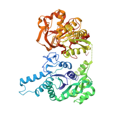
Cryo-EM structures of CTP synthase filaments reveal mechanism of pH-sensitive assembly during budding yeast starvation.
Hansen, J.M., Horowitz, A., Lynch, E.M., Farrell, D.P., Quispe, J., DiMaio, F., Kollman, J.M.(2021) Elife 10
- PubMed: 34734801
- DOI: https://doi.org/10.7554/eLife.73368
- Primary Citation of Related Structures:
7RKH, 7RL0, 7RL5, 7RMC, 7RMF, 7RMK, 7RMO, 7RMV, 7RNL, 7RNR - PubMed Abstract:
Many metabolic enzymes self-assemble into micron-scale filaments to organize and regulate metabolism. The appearance of these assemblies often coincides with large metabolic changes as in development, cancer, and stress. Yeast undergo cytoplasmic acidification upon starvation, triggering the assembly of many metabolic enzymes into filaments. However, it is unclear how these filaments assemble at the molecular level and what their role is in the yeast starvation response. CTP Synthase (CTPS) assembles into metabolic filaments across many species. Here, we characterize in vitro polymerization and investigate in vivo consequences of CTPS assembly in yeast. Cryo-EM structures reveal a pH-sensitive assembly mechanism and highly ordered filament bundles that stabilize an inactive state of the enzyme, features unique to yeast CTPS. Disruption of filaments in cells with non-assembly or pH-insensitive mutations decreases growth rate, reflecting the importance of regulated CTPS filament assembly in homeotstasis.
Organizational Affiliation:
Department of Biochemistry, University of Washington, Seattle, United States.


